Patient recruitment and sample collection
Seminoma samples were provided by Human Genetics Resource Preservation Center of Hubei Province (Department of Biological Repositories, Zhongnan Hospital of Wuhan University), China, a member of International Society for Biological and Environmental Repositories (ID:49623232). All patients have signed the informed consent. Our study followed the ethical rules in accordance with the Declaration of Helsinki and was approved by the ethical reviewing committee of Zhongnan hospital of wuhan university (NO. 2021009K).
RNA extraction and qRT-PCR experiments
RNAs from cell line samples and clinical samples were extracted using RNAiso Plus agent (TaKaRa, Dalian, China) following the standardized protocol. Reverse transcription polymerase chain reaction was performed to generate cDNAs. mRNA expression was detected by qRT-PCR under parameters setting as 95 °C for 25s, 55°C for 30s, 72°C for 90s, with total cycles of 40. Primers used in this study were listed in detail (Supplementary Table 1).
Cell line culturing
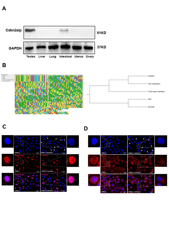
Testicular seminoma tumor cell line (NTERA-2) was acquired from American Type Culture Collection (ATCC, Manassas, VA, USA) on March 10, 2021. RPMI-1640 medium combined with 10% fetal bovine serum (FBS, Hyclone, South Logan, UT, USA). 100 IU/mL penicillin with 100 μg/mL streptomycin (Invitrogen, Carlsbad, CA, USA) were used for culture, with environmental setting under 37°C, with 5% CO2.
Western Blot
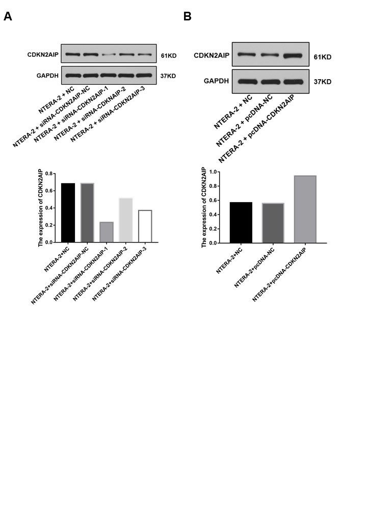
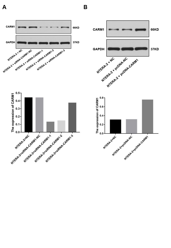
2x10⁶ cells per cell group were firstly washed twice with cold PBS, resuspended and then treated with ice-cold cell lysis buffer RIPA agent (Beyotime, Shanghai, Chian) to extract total protein from the samples. Then BCA protein assay kit was used for sample protein level quantification. Then, protein samples were firstly separated by SDS-PAGE and 10% separating gels and samples were subsequently electroblotted onto PVDF membranes (Immobilon-P, Millipore, Billerica, MA). After blocking in Tris buffer (50 mM Tris, pH 7.5) containing 5% skim milk, the membranes were then incubated overnight at 4°C with primary antibodies. After rinsing with the Tris-Buffered Salin and Tween buffer solution (TBST, Sigma-Aldrich, St. Louis, MO, USA), they were then incubated with the secondary antibody for 2 h. Chemiluminescence was used to expose the protein bands on the membrane. Antibodies used in this study were shown in Supplementary Table 2.
Plasmids
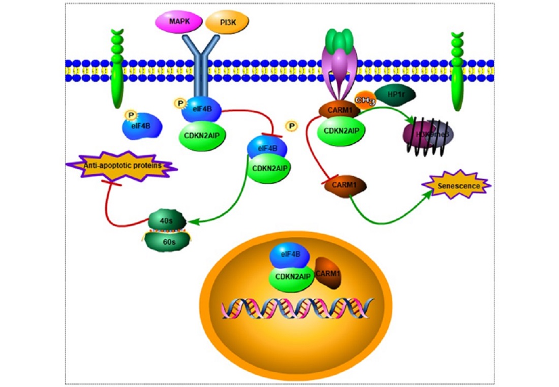
Target genes including CDKN2AIP, CARM1 (Coactivator-associated arginine methyltransferase 1) were cloned by PCR using cDNA as template, then inserted into pcDNA3.1 vector for subsequent transfection
Co-immunoprecipitation (Co-IP)
NTERA-2 cells transfected with tagged CDKN2AIP/CARM1 were cultured in 10-cm dishes. Then cell samples were treated in immunoprecipitation buffer (containing 20 mM Tris-HCl, pH 7.4, 150 mM NaCl, 1 mM EDTA, pH 8.0, 1% NP-40, 1× Protease and Phosphatase Inhibitor). Then 1 μl FLAG/HA antibody or IgG and 10 μl of Protein A/G magnetic beads (pre-washed with lysis buffer) were added and incubated for 5 h under 4 °C condition. Afterwards, the beads were sequentially rinsed with wash buffer. Finally, proteins were eluted from the beads using 2× SDS loading buffer and subsequently treated at 95 °C for 10 min prior to SDS-PAGE and immunoblotting with FLAG and HA antibodies.
Immunofluorescence (IF)
Cells samples were firstly washed with phosphate-buffered saline (PBS), and then paraformaldehyde was used for cell fixation for 15 min. Afterwards, cells were permeabilized with 0.5% NP-40 for 20 min, and then cells were blocked with 5% bovine serum albumin for 30 min. Subsequently, cells were incubated with primary antibody for 2 h at RT, and subsequently incubated with fluorescein conjugated secondary antibody for 1 h. Finally, the DAPI and confocal microscopy were used to stain and image the slides, respectively.
Flow cytometric apoptosis analysis
Apoptosis of NTERA-2 cell groups was analyzed by flow cytometry utilizing Annexin V-fluorescein isothiocyanate (AV-FITC) apoptosis detection kit (640932, BioLegend, San Diego, CA, USA). NTERA-2 cell groups were respectively harvested and further processed according to the standardized procedure for flow cytometric experiment after the initial incubation.
Cellular senescence assay
SA-beta-gal staining assay was performed according to previous description[16]. Each group of cells were stained for 8 hours before evaluation by counting at least 300 cells.
SCID mice xenograft model
Pathogen-free 8–10 weeks old BALB/c SCID mice were used. For testicular seminoma tumor cell inoculation, cells were harvested by trypsination and viable cells (5 x 10⁶) were suspended in 1 ml of cell culture medium. An aliquot of 200 μL of cell suspension was injected subcutaneously for each SCID mouse from every treatment group consisting of 9–11 SCID mice. The mice bearing testicular seminomas were sacrificed when the tumor had reached maximal growth (up to 20% of the body weight of the animal at the beginning of the experiment) or started to ulcerate. Primary tumors were removed, weighed and fixed in 10% PFA in PBS.
Statistical analysis
Statistical analysis was conducted by software package (SPSS 21.0 for Windows, IBM-SPSS Inc., Chicago, IL). Data were presented as the mean SD of three independent experiments. Statistical test of differences between numerical data was performed by standard t-test. Pearson test was conducted to compare gene correlation. P<0.05 was considered to indicate statistical significance.