Study area
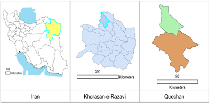
This Cross-Sectional study that was conducted in Quchan city in KhorasanRazavi province in northeastern Iran. It has an area of about 523400 hectares and a population of over 180,000 peoples. The city is located in a mountainous area, elevation 1149 meters above sea level and has cold winters and mild summers. Its rainfall is 200-150mm/year and lies between 37.11° latitude and 58.51 ° E longitude.(Available athttps://www.worldatlas.com/as/ir/30/where-is-quchan.html, https://en.wikipedia.org/wiki/Quchan#Geography). Figure.1 shows ArcGIS geographic location of Iran and Quechan.
Sample collection
Based on statistical advice and previous studies, a total of 296 animals including 168 sheep and 128 goats were sampled from the slaughterhouse in Quechan city for this study. This descriptive cross-sectional study was implemented during the period from August 2016 to April 2017 and samples were collected in four stages in the spring, summer, autumn and winter seasons. According to the seasonal pattern, 65 68, 104 and samples were collected in spring; summer, autumn in winter respectively. Livestock was numbered, randomly selected, whereastheir blood samples(jugular vein) were obtained from a numbered livestock. At the same time, age and gender were also recorded. According to the number of each livestock, about 10 gr of heart and diaphragm tissues were obtained from the same livestock. Totally, we had from each livestock a blood sample, a heart sample and a diaphragm sample. Blood samples were centrifugedin withoutanticoagulant tubes at 8000 rpm for 5-10 minutes and sera were transferred to 1.5ml micro-tubes. All sampling was carried out in compliance with ethical requirements. Till to performing serological and molecular tests, all sera and tissue samples were stored at -20 0C until.
Serological examination
Sera of sheep and goats were examined for anti-toxoplasmagondii antibodies by the modified agglutination test (MAT) (Toxo screen DA, bi-omerieux®, France) as described by Dubey and Desmonts[13].Serumsamples (sheep and goats) were diluted from 1:20 to 1:640. Accordingly to the manufacturer's instructions, antibodies titers of 1:20 or higher were considered positive.
DNA extraction
All positive samples of MAT test were investigated by PCR assay on the heart and diaphragm of the same animals. For this purpose, 20 mg of tissues were transferred into a sterile plate and crushed. This work continued until the sample was completely homogeneous. Then, homogenized tissue is transferred into a 1.5 ml microcentrifuge tube and subsequent stages of DNA extraction were performed using (Gene All, Exgene,Cell SV mini, Korea) kit and according to the manufacture`s instruction.
Nested-PCR for B1 gene
The Nested-PCR assays were accomplished based on two repeated genomic targets, B1, to detect T. gondii DNA in contaminated tissues. B1 Gene is 35 times reported and has high sensitivity and specificity of PCR in the determination of the contamination of clinical samples with T.gondii[13] .Two PCR primer pairs of the B1 gene, S1 (5´-CGACAGAAAGGGAGCAAGAG-3´) and AS1 (5´-ACGCTGTGTCTCCTCTAGGC-3´), S2 (5´-TCTTCCCAGACGTGGATTTC-3´) and AS2 (5´-CTCGACAATACGCTGCTTGA-3´), eventually amplifying a 531 bp fragment were used. The first amplification was carried out in 20 µl of reaction mixture containing 1 µl of each primer (S1 and AS1), 10µl Master mix (Ampliqon Company, Denmark), 2 µl extracted DNA from heart or diaphragm samples and 6µl Distilled water sterilized. The first PCR was performed in a thermocycler (Flex Cycler) for initial denaturation at 94 C for 3 min, this step was followed by 35 cycles of denaturation at 94 C for 30 s, annealing at 60 C for 30 s, extension at 72 C for 2 min and a final extension step at 30 C for 1 min. The second amplification was performed in 20 µl reaction mixture. The first PCR product was diluted with a ratio of 1:40 to distilled water, and then used as a template. Twenty µl reaction mixture was containing 1 µl of each primer (S2 and AS2), 8µl Master mix (Ampliqon Company, Denmark), 1 µl of our new template and 9µl distilled water sterilized. The second PCR was performed in 30 cycles.The PCR products were electrophoresed in a 1.5% Agarosegel in tris-borate-EDTA 0.5X (TBE 0.5X) buffer and stained with Ethidium bromide. Additionally,negative and positive control respectively include sterile waterand extracted DNA from T. gondiitachyzoites RH-strain was used in this method.
Nested PCR for GRA6 gene
The positive samples of Nested-PCR of B1 gene included in analyzing by Nested-PCR of GRA6 gene. GRA6, ahighly polymorphicgene is repeated in the genome of the T. gondii. This gene is suited to distinguish between three typesI, II and III from each other, especially type III which is close to type I. Two PCR primer pairs of the GRA6 gene, GRA6FO (5´GGCAAACAAAACGAAGTG-3´) and GRA6RO (5´-CGACTACAAGACATAGAGTG-3´) used in first amplification, and GRA6R (5´-GTAGCGTGCTTGTTGGCGAC-3´) and GRA6 (5´TACAAGACATAGAGTGCCCC-3´) used in second amplification.The first amplification was carried out in 25 µl of reaction mixture containing 1 µl of each primer (GRA6FO and GRA6RO), 8µl Master mix (Ampliqon Company, Denmark), 5µl extracted DNA of heart or diaphragm samples and 10µl Distilled water sterilized. The first PCR was performed in a thermocycler (Flex Cycler) for initial denaturation at 94 C for 5 min, this step was followed by 35 cycles of denaturation at 94 C for 30 s, annealing at 54 C for 60 s, extension at 72 C for 90 s and a final extension step at 72 C for 7 min[14].The second amplification was performed in 25 µl reaction mixture. The first PCR product used as a template while diluted with a ratio of 1:10 to distilled water.Twenty-five microlitresreaction mixture was containing 1 µl of each primer (GRA6R and GRA6), 8µl Master mix, 1µl of our new template and 14µl Distilled water sterilized. The second PCR was performed at the annealing temperature of 60 C for the 60s[15]. The PCR products were electrophoresed in a 1.5%agarosegel in tris-borate-EDTA 0.5X (TBE 0.5X) buffer and stained with ethidium bromide.To differentiate the three types (І, II, III) of T.gondii,all positive samples of Nested PCR for GRA6 gene were used to performing PCR-RFLP technique.
PCR-RFLP
The GRA6 gene amplified product was digested with MseI with MseIrestriction endonuclease (10 U/μl, 300 units), (Fermentas, Thermo Scientific, USA). As described by the manufacturer, 15 ml of PCRproduct was exposedto 1.5 U of MseIen-zyme and 2 U buffer R and incubated at 65 °C for 4 h. The restriction fragments were separated by electrophoresis in 2% agarose gel followed by staining with ethidium bromide and visualization under UV. The cut position of MseIin GRA6 genes of types I, II, and III was 168 bp and 712 bp, 71 bp and 694 bp, and 71 bp, 168 bp, and 712 bp, respectively.
Sequencing
The GRA6 gene amplified product (with suitable quality in PCR-RFLP) sent to Macrogen company (South Korea) to sequence analyzing and also to obtain more accurate results from the genotype of the T. gondii(I, II, III). Results were aligned with BioEdit and sequence Scanner program and compared to the following sequence data available from GeneBank: AJ635332, AF239283, AF239292 and AF239284.The maximum-likelihood analysis was employed to estimated phylogenetic relationshipsamong genotypes. Additionally, Mega6 andBioEdit software were used to construct thephylogeny tree to compare our collected isolates against types submitted in Genebank as well as to demonstrate homology of obtained sequences respectively.
Statistical Analysis
Differences in T. gondii prevalence with variables such as season, sex and age and calculate the prevalence rate was analyzed using Pearson Chi-square test and crosstab. Statistical analysis was performed using SPSS version 23 software for Windows.The p-values less than 0.05 were considered as statistically significant.