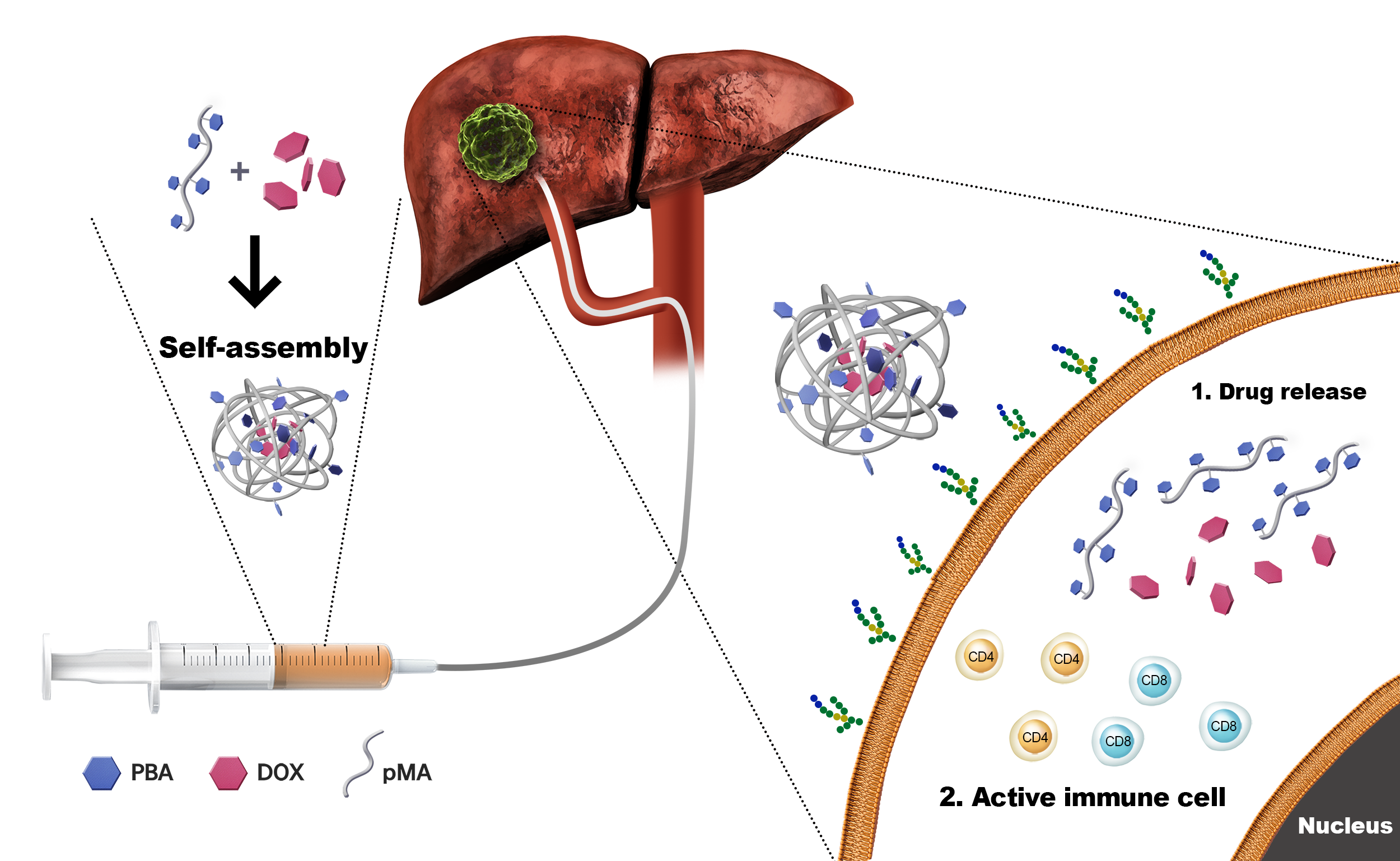
In the present study, we demonstrated that there is an effective anticancer effect and immune response in liver cancer through TACE using pPBA-Dox nanocomplexes.
According to the BCLC guidelines, many HCC patients attempt LRT as the primary treatment method, and 40% of HCC patients attempt TACE as their primary treatment (21). However, there is a significant difference in tumor burden and liver function depending on the patient, making it difficult to achieve the same effect through TACE. Accordingly, systemic chemotherapy has been proposed, and a new paradigm for anticancer treatment has been proposed with the recent development of immune checkpoint inhibitors (ICIs). However, due to the low response rate of ICIs, clinical studies of anticancer treatment through combination treatment with existing treatment methods are actively progressing (22–25).
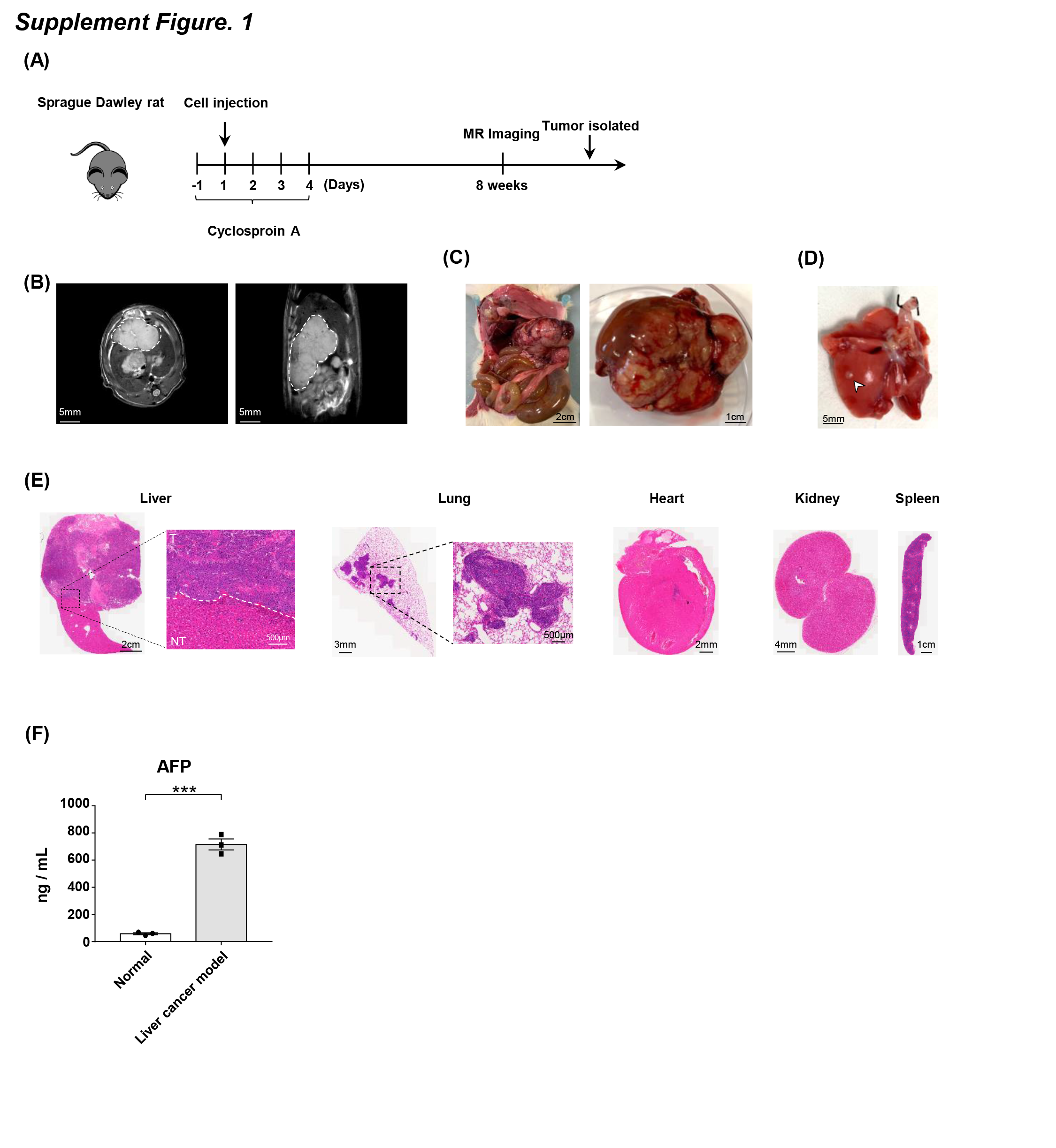
We verified the anticancer effect of liver cancer through TACE of pPBA-Dox nanocomplexes and confirmed the change in immune cells in the liver according to drug infusion through TACE. To confirm this hypothesis, we established an animal model of TACE. Generally, the rabbit liver cancer model using VX2 tumor cells has been widely used because the size of the rabbit hepatic artery is appropriate for the introduction of microcatheters. The rabbit model has the advantage of being easy to perform angiography with large blood vessels, but it is difficult to handle, and as neoplasm VX2 tumor cells are not hepatocyte-derived tumors, there is a limitation in that they are physiologically different from HCC (26, 27), so this model differs from liver cancer in humans (28). For these reasons, there is a need for rodent HCC models derived from hepatocellular carcinoma appropriate not only for TACE but also for various studies of anticancer therapies.
Rat liver cancer models using two rodent HCC cell lines are widely used. One is a Morris hepatoma model using McA-RH7777 hepatoma cells in Buffalo rats, and the other is a Novikoff hepatoma model using N1-S1 hepatoma cells in SD rats. Although McA-RH7777 cells have a higher tumor induction rate than N1-S1 cells, buffalo rats are not widely available in some countries and are generally more expensive than SD rats. Therefore, an HCC model was established in the widely used SD rats using McA-RH7777 cells with high tumorigenicity, and TACE was attempted using this model (29). The SD rats showed a tumor formation rate of greater than approximately 95%, and the tumor size increased by 14.29 ± 2.78 mm and 662.3 ± 86.28 mm3 2 weeks after cell injection. In addition, ALT and AFP were also significantly increased. Interestingly, approximately 8 weeks after cell injection, metastasis to the lung was also observed, showing the possibility of this model as a late metastasis model.
After TACE, tumor size and serum analysis according to each drug showed that the pPBA-Dox nanocomplex group had a higher anticancer effect than the Dox group. The tumor size of the pPBA-Dox nanocomplex group decreased more than that of the Dox group. For AFP, a biomarker of HCC, the AFP reduction ratio in the pPBA-Dox nanocomplex groups was − 0.02 ± 0.03, which was significantly reduced compared to the other groups in which AFP was increased. Both ALT and creatinine were within the normal range, suggesting significant results through hematological analysis. Through these results, it was confirmed that pPBA-Dox nanocomplex infusion through TACE showed a high anticancer effect on liver cancer but did not show hepatotoxicity or nephrotoxicity.
After TACE in the pPBA-Dox nanocomplexes and Dox groups, it was confirmed through H&E staining that tumor necrosis occurred and immune cells infiltrated around the tumor. In addition, there is a report that PBA used as a drug delivery agent also affects the proliferation of PBMCs (30). Therefore, we confirmed the change in the T cell population in the liver after TACE to confirm the possibility of ICIs after TACE. Because of the heterogeneity of liver cancer, it is difficult to achieve a high therapeutic effect with monotherapy, and to overcome this, combination therapy using several treatment methods is being attempted (31, 32). Recently, in many clinical trials, studies of combination therapy using LRT and ICIs have been conducted (33). It is difficult to obtain patient liver tissue after TACE, and some results have confirmed T cell function after radiofrequency in a murine xenograft model, but there is no result confirming any changes in the T cell population and function in the liver after LRT (34). Other than that, there are only studies indirectly confirming changes in the T cell population and function after LRT in PBMCs (23, 35–37). Therefore, this study was able to directly confirm the possibility of ICIs after LRT, especially TACE, by confirming a change in the T cell population in the liver after LRT.
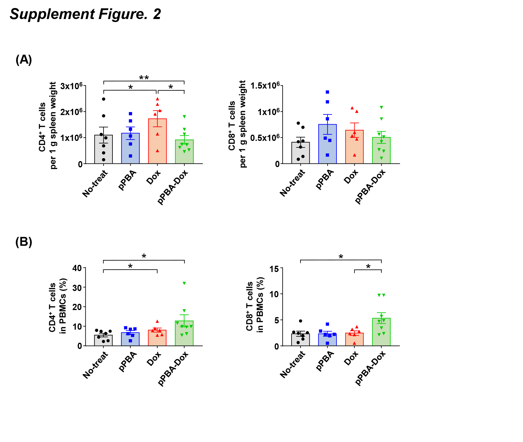
CD4+ and CD8+ T cells were significantly increased in the pPBA-Dox nanocomplex group compared with the Dox group. As a hypothesis to explain the increase in the immune cells in the pPBA-Dox nanocomplex groups, we suggest that pPBA-Dox nanocomplexes accumulate and remain in tumor cells for a long time, as shown in previous studies (15, 16), which promotes immune modulation and additionally induces necrosis of tumor cells. These effects induce immunogenic cell death (ICD), and dying tumor cells upregulate and release death-associated molecular patterns (DAMPs), such as calreticulin (CRT), adenosine triphosphate (ATP), and high-mobility-group Box 1 (HMGB1) (38). Thereafter, phagocytes are recruited, and the activation of dendritic cells (DCs) loads major histocompatibility complex (MHC) molecules, which eventually act as antigens on T cells and enhance T cell infiltration (39). This promotes the cytotoxic T cell immune response, which promotes tumor death.
In the rat model, the pPBA-Dox nanocomplex group had more CD4+ and CD8+ T cells than the Dox group after TACE, which means that T cell infiltration was higher in the pPBA-Dox nanocomplex group than in the Dox group. These results suggest that pPBA-Dox nanocomplexes induce more cell death than Dox through tumor-specific targets, thereby promoting T cell infiltration. On the other hand, T cell infiltration did not increase in the pPBA group. These results indicate that the drug delivery nanomaterial pPBA, a foreign material, does not induce an immune response in the liver.
After the pPBA-Dox nanocomplexes were infused via TACE, the CD4+ and CD8+ T cell populations increased, and the T cells were activated. After TACE, the expression of CD69, Ki-67 and PD-1 in the CD8+ T cells was increased in the group infused with pPBA-Dox nanocomplexes in the liver. In addition, the expression of CD69 and Ki-67 in CD4+ T cells was high in the pPBA-Dox nanocomplexes. This suggests that activated CD8+ T cells secrete cytokines and cytolytic molecules to kill tumor cells, and activated CD4+ T cells assist cytotoxic T cells and other immune cells in performing effector functions, thereby exerting anticancer effects (40–44). These results demonstrated that when pPBA-Dox nanocomplexes were infused through TACE, they affected immune cells and increased the anticancer effect. Similar to the results of the cytotoxicity assay, immune cells isolated from the pPBA-Dox nanocomplex group exhibited higher anticancer effects on tumor cells. During the cytotoxicity assay process, PHA is a nonspecific stimulant of normal lymphocytes and has the disadvantage of stimulating not only cytotoxic T cells but also all immune cells, such as NK cells. This needs to be confirmed in a future study, but there may have been a change in the frequency of immune cells depending on each drug after TACE. Therefore, even if the same stimulation was given to each group with PHA, it is expected that pPBA-Dox nanocomplexes would have a strong cytotoxicity effect.
In the present study, the pPBA-Dox nanocomplexes had a strong anticancer effect when treating liver cancer using TACE and they induced changes in the immune cell population in the liver. This change in immune cells promoted the infiltration and activation of CD4+ and CD8+ T cells in the liver. In conclusion, our study suggests a targeted cancer therapy strategy using pPBA-Dox nanocomplexes is effective for the treatment of HCC, and it may be a promising candidate anticancer drug.