Animal models
The study was approved by the Institutional Animal Care and Use Committee of Tongji Medical College, Huazhong University of Science and Technology. Adult female Sprague–Dawley (SD) rats (8 weeks of age) were purchased from Spelford Biotechnology Co. (Beijing, China). The experiments were performed after a 1–2-week period of adaptive feeding, during which each rat was provided with chow and fresh water under the same control conditions (21 ± 3°C, 12-h/12-h light/dark cycle, lights on at 8:00 a.m.). The experimental study involving three groups: the IUA group, sham-operated group and blank control group. For animals in the IUA and sham groups, the right uterine horn was subjected to double damage or sham surgery (IUA-R or Sham-R, respectively) during the estrous phase, and the left uterine horn was used as a control (IUA-L and Sham-L, respectively). The rats were anaesthetised with sodium pentobarbital and placed in the supine position. The abdomen was shaved and disinfected three times with iodophor, and a transverse incision of approximately 3 cm was made on the lower abdomen. The procedure was performed with reference to the method described by Li et al.31, 32. In brief, IUA was modelled by mechanical and infectious injury. To create a mechanical injury, the endometrium was scratched with a spatula to abrade the surface of the uterine cavity. To create an infection injury, bacterial lipopolysaccharide (LPS) surgical cotton thread was left in the uterine cavity; the end of the surgical thread was left in the abdomen, and the abdominal incision was sutured. After 48 h, the tail wire was gently pulled, and the LPS surgical cotton thread was removed from the uterus. In the sham-operated group, the surgical incision was made and sutured without infection injury. This method of modelling produces a more clinically appropriate representation of IUA. The animals were euthanised in batches on days 3, 7, 14 and 28.
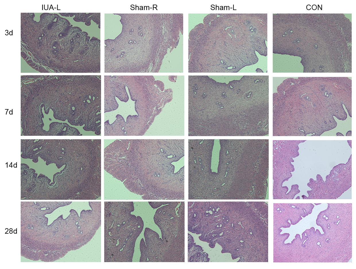
HE staining
The uterus was fixed in 4% paraformaldehyde for 24 hours and then embedded in paraffin. Paraffin-embedded tissue sections were cut, dewaxed and hydrated, and stained using an HE dye set (G1003, Wuhan Servicebio Technology Co., Ltd., China). The stained sections were observed under a light microscope (Olympus, Japan). The number of glands was counted and IUA was evaluated in images of the sections.
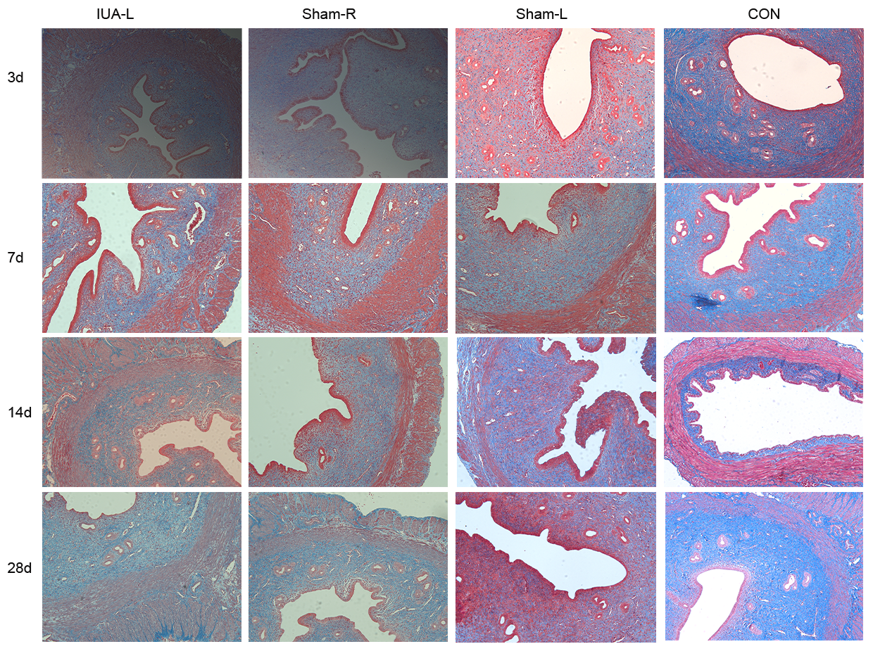
Masson staining
Paraffin-embedded tissue sections were dewaxed, hydrated, and stained using a Masson dye kit (G1006, Wuhan Servicebio Technology Co., Ltd.). The sections were observed under an optical microscope (Olympus) to identify blue-stained collagen fibres, and the area of labelled collagen fibres relative to the overall field of view was calculated using ImageJ software (National Institutes of Health, USA).
Immunofluorescence
Immunofluorescence staining with antibodies specific for α-SMA (A7248, ABclonal; dilution, 1:1,00), vimentin (A19607, ABclonal, dilution, 1:1,00) and CD31 (ab182981, Abcam; dilution, 1:1,000) was performed. In brief, paraffin-embedded tissue sections were dewaxed and hydrated. After antigen retrieval, the tissues were subjected to autofluorescence quenching and incubated in serum to block nonspecific antibody binding. The tissue sections were placed on slides and incubated with the above-listed primary antibodies at 4°C overnight. Subsequently, the sections were incubated with a CY3-conjugated goat anti-rabbit secondary antibody (Servicebio; dilution, 1:300), and the nuclei were counterstained with DAPI (C0065, Beijing Solarbio Science & Technology Co., Ltd.).
Immunohistochemistry
Paraffin-embedded tissue sections were dewaxed and hydrated. After antigen retrieval, endogenous peroxidase activity was blocked by incubating the tissues in 3% hydrogen peroxide, followed by blocking in rabbit serum for 30 min to inhibit nonspecific antibody binding. The tissues were placed on slides and incubated with primary antibodies overnight at 4°C, followed by a 1-h incubation with a goat anti-rabbit secondary antibody (D3002, Long Island Antibody, China; dilution, 1:1,000). Subsequently, the slides were stained using a 3,3′-diaminobenzidine colour development kit (DA1010, Beijing Solarbio Science & Technology Co., Ltd.) at room temperature. Finally, the nuclei were counterstained with haematoxylin, and the samples were dehydrated and covered with glass coverslips.
Real-time polymerase chain reaction (qPCR) analysis
Excised rat uterine tissues were homogenised in the presence of TRIzol reagent (Invitrogen, USA) according to the manufacturer’s instructions. After determining the purity and concentration, RNA was reverse-transcribed to single-strand cDNA. Real-time quantitative PCR analysis was performed using a SYBR Green PCR Kit (Yeasen Biotech Co., Ltd., China) in a total reaction volume of 10 µL, including 0.5 µL of cDNA, 5 µL of Master Mix, 1 µL of forward and reverse primers and 3.5 µL of distilled H2O. The 2-ΔΔCt method was used to determine relative gene expression. The following primers were used to detect mRNAs encoding proteins of interest: CD31, 5′-GGT AAT AGC CCC GGT GGA TG-3′ (forward) and 5′- TTC TTC GTG GAA GGG TCT GC-3′ (reverse); α-SMA: 5′-ACC ATC GGG AAT GAA CGC TT-3′ (forward) and 5′-CTG TCA GCA ATG CCT GGG TA-3′ (reverse); vimentin: 5′-CGA GTT CAA GAA CAC CCG CA-3′ (forward) and 5′- GCG CAC CTT GTC GAT GTA GT -3′ (reverse). As an endogenous control, the reference gene GAPDH (forward primer, 5′-AGT GCC AGC CTC GTC TCA TA-3′; reverse primer, 5′-GGT AAC CAG GCG TCC GAT AC-3′) was amplified to enable the normalisation and relative quantitative analysis of the expression of genes encoding CD31, α-SMA and vimentin . The following conditions were applied to the reactions using a thermocycler (Quantagene q225 real-time PCR system, Novogene, China): 40 cycles at 95°C for 5 s, 60°C for 20 s and 72°C for 20 s. All reactions were performed at least three times.
Western blot analysis
Total proteins were extracted from rat uterine tissues using radioimmunoprecipitation assay buffer (Beyotime, China) and phenylmethylsulphonyl fluoride (Beyotime, China). The total protein concentration in each resulting lysate was quantified using a bicinchoninic acid assay kit (Beyotime, China). Next, 40 µg of total proteins from each sample were loaded onto a 10% sodium dodecyl sulphate–polyacrylamide gel, subjected to electrophoresis to separate the proteins, and transferred to polyvinylidene fluoride membranes (Millipore, Germany). The membranes were incubated in a 5% non-fat milk solution to block nonspecific antibody binding and incubated overnight at 4°C with primary antibodies specific for the following proteins: α-SMA (A7248, ABclonal; dilution, 1:1,000), vimentin (A19607, ABclonal; dilution, 1:1,000) CD31 (A2104, ABclonal; dilution, 1:1,000) and GAPDH (60004-1-lg, mouse monoclonal, Proteintech; dilution, 1:4000). After three 10-min washes with Tris-buffered saline containing Tween-20, the membranes were incubated with appropriate horseradish peroxidase-conjugated secondary antibodies (ABclonal, China). Specific proteins were detected by treating the blots with enhanced chemiluminescence reagents (Biosharp, China) and exposing them to film. The protein expression levels were analysed quantitatively using AlphaEaseFC software (Alpha Innotech, USA), and the protein levels were normalised against the level of GAPDH in each sample.
Statistical analysis
All measurements are shown as means ± standard deviations, and each set of data was derived from at least three independent experimental replications. Data from different groups were analysed using t tests and one-way ANOVAs, followed by post hoc comparisons using Dunnett’s test. SPSS 23.0 software (v.23.0; SPSS Inc., Chicago, IL, USA) was used for data processing and statistical analysis. GraphPad Prism 8.0 software (v.8.0; GraphPad Software Inc., San Diego, CA, USA) was used to create and process the histograms. A p value <0.05 was considered to indicate a statistically significant difference.