Patient samples
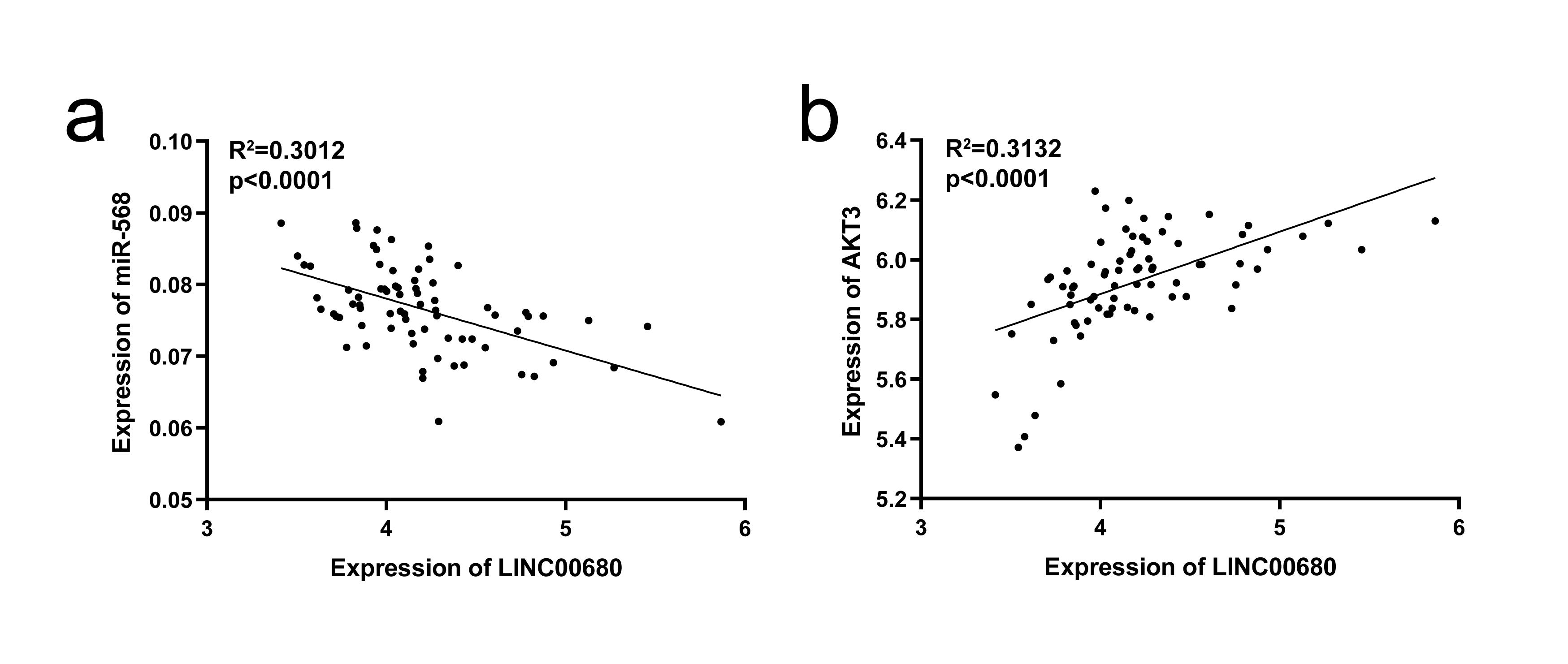
A total of 74 pairs of HCC cancerous and corresponding non-cancerous tissues were gathered from HCC patients who underwent surgery from 2017 to 2020 at The First Affiliated Hospital of Guangxi Medical University. These tissue samples were immediately transferred into liquid nitrogen right after the surgery and then stored at -80 ºC for future use. This study was reviewed and approved by the ethics committee of The First Affiliated Hospital of Guangxi Medical University, and written informed consent in accordance with the Declaration of Helsinki and its later revision was provided by all the patients.
Cell lines and cell culture
HCC cell lines SNU-449, SNU-182, Huh7, LM3, Bel-7405, SK-hep1, Hep3B and normal human liver cell line L02 were purchased from the American Type Culture Collection (Manassas, VA, USA) or Institute of Biochemistry and Cell Biology (Chinese Academy of Sciences, Shanghai, China). SNU-449, SK-hep1 and SNU-182 were cultured in RPMI-1640 medium (Gibco, USA) containing 10% fetal bovine serum (FBS, Gibco). The other cells were cultured in high-glucose Dulbecco’s modified Eagle medium (DMEM) (Gibco) containing 10% FBS. Penicillin (100 U/ml) and streptomycin (100 μg/ml) were supplemented to the medium to reduce the chance of microbial contamination. All the cells were maintained in a humidified atmosphere at 37℃ with 5% CO2/95% air.
Cell transfection
Short hairpin RNA (shRNA) for LINC00680 knockdown and its negative control were obtained from GeneChem (Shanghai, China). pcDNA3.1 (+) vector for LINC00680 overexpression and its corresponding control, miR-568 mimics, miR-568 inhibitor and their negative controls, AKT3-targeted siRNA, pcDNA3.1 (+) vector for AKT3 overexpression, and their corresponding negative controls were all synthesized by GenePharma (Shanghai, China). Cells were seeded on a six-well plate at a density of 5 × 10⁵ cells /well. Transfection operation began after 24 h incubation at 37 ºC in a humidified incubator. All the in vitro transfections were performed using Lipofectamine TM 3000 Reagent (Thermo Fisher Scientific) according to the manufacturer’s instructions. Lentiviral infection method was used to deliver sh-LINC00680 into HCC cell lines used for in vivo mouse xenograft tumor model.
Quantitative real time polymerase chain reaction(qRT-PCR)
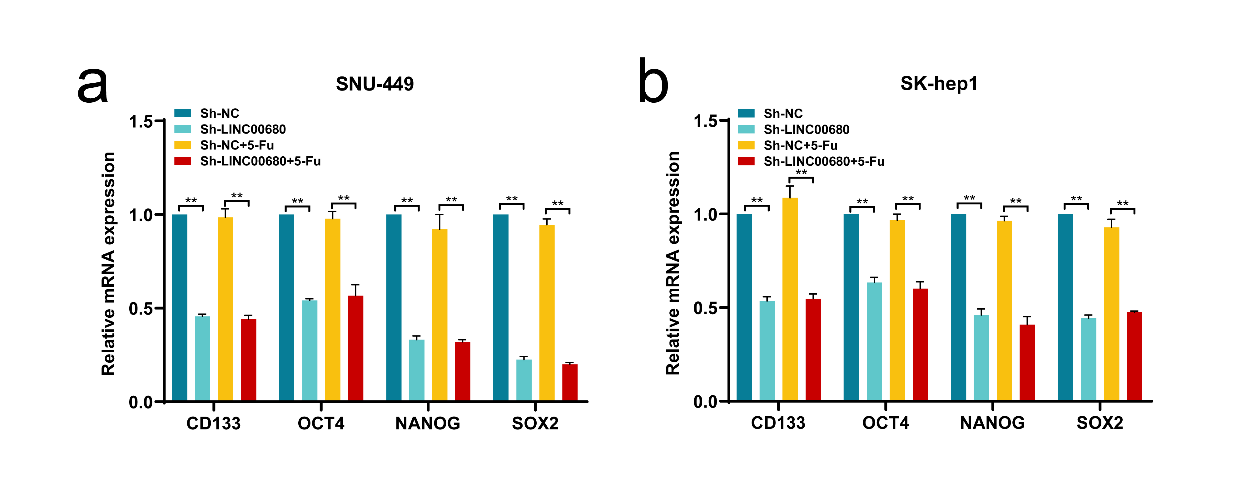
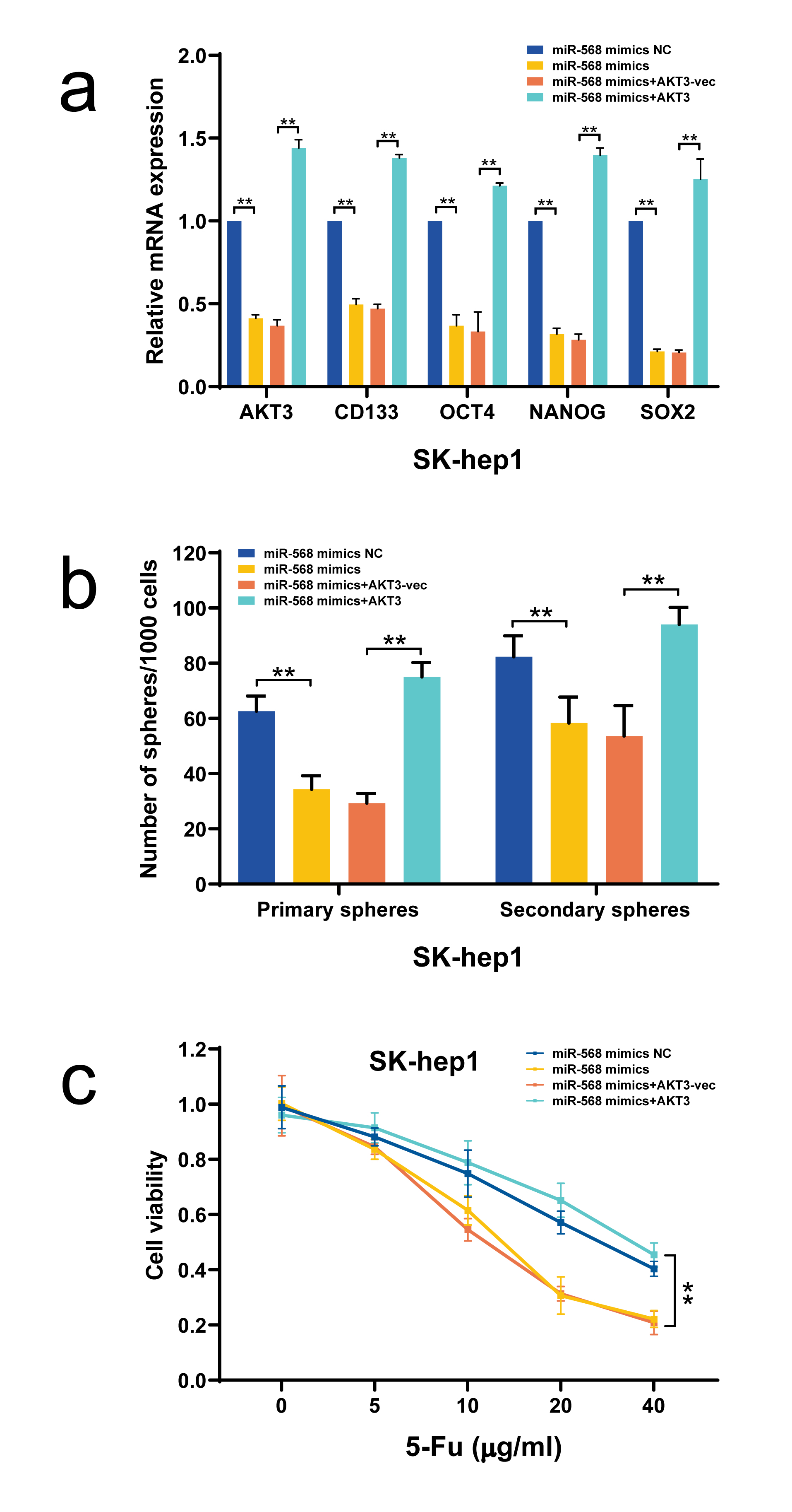
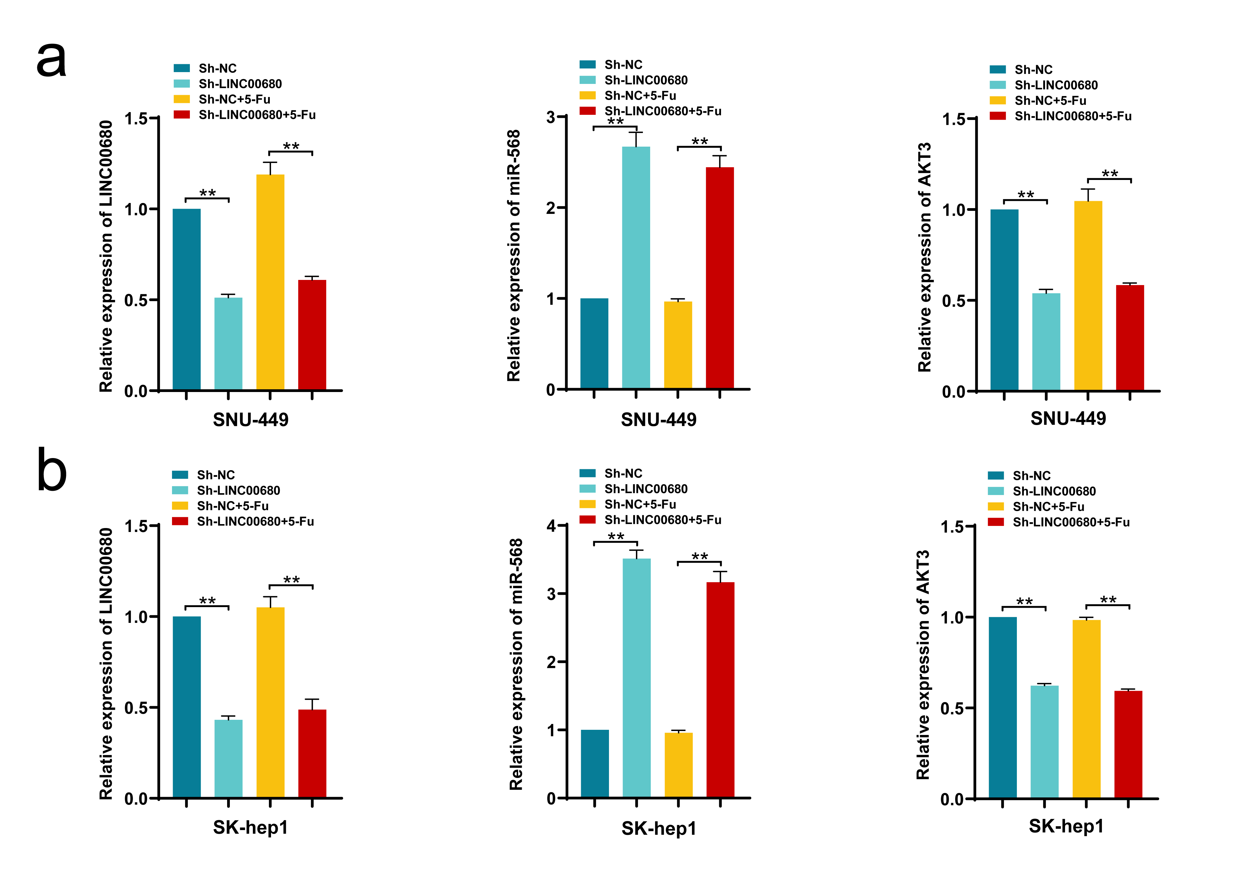
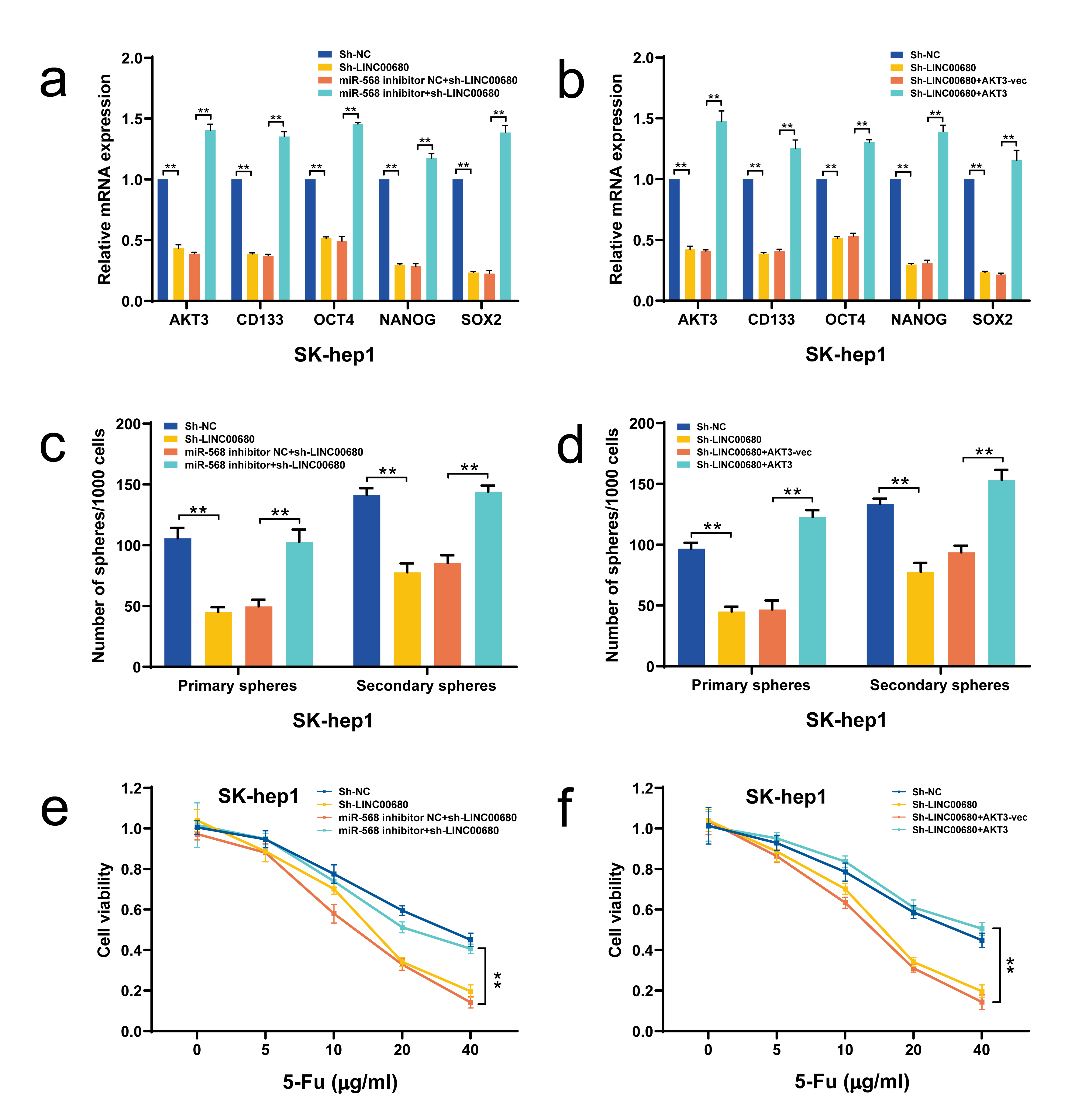
Total RNA were extracted from cells and tissues by RNA simple Total RNA Kit (TIANGEN, DP419). The reverse transcription of cDNA was carried out by Revert Aid First Strand cDNA Synthesis Kit (Thermo Scientific, #K1622) and poly (A) polymerase Reaction Buffer (NEB, M0276s) according to the manufacturer’s instructions. QRT-PCR was performed by iTaqTM Universal SYBRâ Green Supermix. The fold change of RNA expression was quantified using the 2-ΔΔCt method after normalizing with GAPDH or U6 expression. The primers used to detect miR-568 were designed by GeneCopoeia with their sequences protected by a patent (http://www.igenebio.com/tech/datasheet/index.php). The mRNA Primers used are listed in Supplementary Table 1.
Western blotting
Protein samples were extracted using cold RIPA lysis buffer containing protease inhibitor cocktail (Roche, Mannheim, Germany), with protein concentration determined by the BCA method. Equal amounts of protein lysates were run on a sodium dodecyl sulfate polyacrylamide gel electrophoresis (SDS-PAGE) Bio-Rad system. Separated proteins were detected with the corresponding antibodies and visualized using enhanced chemiluminescence (ECL). The primary antibodies included those against AKT3, p-AKT3, mTOR, p-mTOR, elF4EBP1, p-elF4EBP1, p70S6K, p-p70S6K, CD133, OCT4, NANOG, SOX-2 and β-actin. All these antibodies were purchased from Abcam Corporation (Cambridge, MA, USA).
Sphere formation assay
The Cells in exponential growth phase were seeded in ultralow attachment six well plates (Corning) at a density of 1000 cells/well and cultured for one week (Primary spheres) in DMEM/F12 medium (Invitrogen, Shanghai, China) supplemented with B27 (1:50, GIBCO, Shanghai, China), 20 ng/ml EGF (Sigma, Shanghai, China), and 20ng/ml bFGF (Sigma, Shanghai, China). Subsequently, the primary spheres were collected, dissociated with trypsin and plated again to generate secondary spheres through the same process. Finally, the number of the spheres was counted a microscope.
Cell proliferation assay
Cell count kit-8 (CCK8, Dojindo, Japan) and colony formation assay were used to determine cell proliferation. For CCK8 assay, cells were seeded in 96-well plates at a density of 1 × 10³ cells/well, grew overnight, and the treated with different concentration of 5-Fu for 48 h. Subsequently, 10 μl CCK-8 solution was added to each well and incubated for another 2 h at 37 ℃ under a humidified 5% CO2/95% air atmosphere. Absorbance values were measured on a microplate reader (spectramax plus384, Molecular Devices, USA) at 450 nm. For the colony-formation assay, cells were seeded in 6-well plates at a density of 1000 cells/well, and grew for 7 days with or without 5-Fu. After rinsed twice by PBS, formed cell colonies were fixed in 70% methanol for 30 min and stained with 0.5% crystal violet for 30 min. The cell colonies larger than 50 cells were counted for comparison between groups.
Analysis of the tumor-forming potential in vivo
6-week-old male BALB/c nude mice (Shanghai Slac Laboratory Animal Co. Ltd., China) were obtained and bred under a SPF (specific pathogen-free) condition. LINC00680 stably downregulated SNU-449 and SK-hep1 cells and their negative controls were subcutaneously injected into the mice (5 mice/group). The volume of tumors was continuously measured using a Vernier caliper. Five weeks later, the mice were euthanatized to measure the tumor weight. The animal experimental protocols were in accordance with institutional guidelines approved by the Animal Care and Use Committee.
Dual luciferase reporter assay
The sequences of wild- or mutant-type AKT3 3'-UTR were inserted into PmiRGLO dural-luciferase reporters. Thereafter, the recombinant plasmids were co-transfected with miR-568 mimics or miRNA mimics negative control (NC) into 293T cells by lipofectamine TM 3000 (Thermo Fisher). After 36 h transfection, Luciferase assay system (Promega, Madison, USA) was used to determine the relative luciferase activity normalizing to renilla luciferase activity. The binding between LINC00680 and miR-568 was verified using a similar method.
RNA immunoprecipitation (RIP) assay
We used Magna RIP™ RNA-Binding Protein Immunoprecipitation Kit (Millipore, USA) to perform RIP experiments according to the manufacturer’s instructions. Beads were incubated with AGO2 antibodies and washed with wash buffer, the complexes were then incubated with 0.1% SDS/Proteinase K (0.5 mg/ml, 30 min at 55 ℃) to remove proteins, LINC00680 and miR-568 were detected by qRT-PCR. The supernatant of RIP lysate was used to test the expression of AGO2 by western blot.
RNA pull-down assay
Pierce™ RNA 3' End Desthiobiotinylation Kit(Thermo, 20163)was used to label LINC00680 for attachment to streptavidin magnetic beads which can capture protein complex combined with labeled LINC00680. According to the manufacturer’s instructions, Thermo Scientific Pierce Magnetic RNA-Protein Pull-Down Kit (Thermo, 20164) was used to perform RNA pull-down assay. AGO2 was assessed by western blot, and LINC00680 and miR-568 were detected by qRT-PCR.
Microarray analysis
We performed a microRNA microarray analysis using SNU-449 cells transfected with miR-568 mimics or its negative control. Total RNA were extracted by RNA simple Total RNA Kit (TIANGEN, DP419) according to the manufacturer’s instructions. NanoDrop one (Thermo Fisher Scientific) was used for measurement of RNA quantity. Microarray analysis were performed by Huayin Health Medical Group Co, Ltd (Guangzhou, China). These obtained data were then subjected to KEGG and GO analysis. Cluster 3.0 and Java Tree View software were used to visualize heat maps.
Statistical analysis
Data were presented as the mean ± standard deviation (SD) from at least three independent experiments and analyzed using SPSS 24.0 for Windows (SPSS, Chicago, IL, USA). Student’s t test or one-way analysis of variance (ANOVA) plus Tukey’s post-hoc test was used for comparisons between groups. Kaplan-Meier method was used for analysis of overall survival (OS) and disease-free survival (DFS). P-values <0.01 were considered statistically significant.