Materials
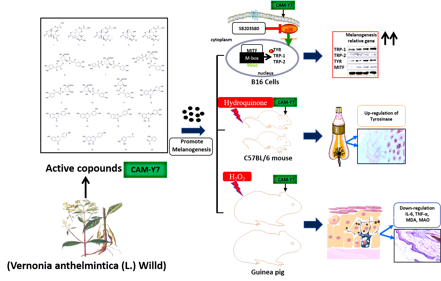
CAM-Y7 was prepared by The Key Laboratory of Plant Resource Sand Chemistry of Arid Zone, Xinjiang Technical Institute of Physics and Chemistry, Chinese Academy of Sciences: One kilo V. anthelmintica seeds was powdered and extracted twice using 12 liters of 80% ethanol/water at 80°C, each extraction lasting 2h. Both extracts were pooled and filtered, and the filtrate was concentrated in a rotary vacuum evaporator to 0.1 g/m. The crude drug was then adsorbed on 2.5 L macroporous HPD300 resin at the flow rate of 2-bed volume per hour (BV/h), purified with 3BV of water, and then eluted with 3BV of 60% ethanol/water. The eluent was evaporated under reduced pressure and dried at 45°C to obtain 62g of the purified drug powder. And dissolved in DMSO and stored at -20°C as a stock solution (10 mg/ml). L-DOPA (CAS No 59-92-7) was obtained from Generay Biotech (Shanghai, China), 8-MOP (CAS:298-81-7) was purchased from Sigma Aldrich (Milan, Italy), CCK-8 was obtained from Trans Gen Biotech (Beijing, China), RIPA Lysis buffer (AR0105-100) was obtained from BOSTER Biological Technology (Wuhan, China), BCA kit assay (PP02) was purchased from Biomed Biological Technology (Beijing, China). Antibodies against β-actin (8H10D10) and Tyrosinase (C-19), TRP-1 (H-90), TRP-2 (H-150) were purchased from Santa Cruz Biotechnology Inc. (Santa Cruz, CA, USA). Anti-MITF antibody was obtained from Abcam (Cambridge, UK). Goat anti-rabbit (BA1054), p38 (L53F8), p-p38 (Thr180/Tyr182) (28B10), SAPK/JNK (#9252s), p-JNK (Thr183/Tyr185), ERK(L34F12), and p-ERK(Thr202/Tyr204) (E10) were purchased from Cell Signaling Technology (Danvers, MA, USA), goat anti-mouse (BA1050), rabbit anti-goat (BA1060) antibodies were obtained from BOSTER Biological Technology (Wuhan, China), interleukin 6 (IL-6), tumor necrosis factor-α (TNF-α), Tyrosinase (TYR), and monoamine oxidase (MAO) and malondialdehyde (MDA) specific ELISA kits were obtained from Elabscience Biotechnology (NJJCBIO Co., Nanjing, China).
Drug analysis
Fifty milligrams of the sample powder were dissolved in 10 ml methanol and filtered through 0.22μm nylon membrane microfilters (Shimadzu-GL, Japan) and chromatographically analyzed using a Shimadzu LC-2030c HPLC fitted with a photodiode detector (PDA) (Shimadzu-GL, Japan) (Fig. 1A). Reversed-phase separation was performed on a Shimadzu Shim-Pack GIST C18 column (4.6 × 250 mm, 5 μm, Shimadzu-GL, Japan) at 35°C. The mobile phases consisted of (A) 0.1% phosphoric acid in water and (B) acetonitrile. The sample was injected (5μl injection volume) onto the column and eluted at 1 ml/min flow. The fractions were detected using U.V. light at 210 nm, 254 nm, and 330 nm. Quantitative analysis was performed using HPLC.
Cell culture
Experiments were conducted in B16 cells (B16, Cat# TCM2), which were cultured in DMEM (Gibco Life Technologies, Waltham, MA, USA) supplemented with 10% FBS (Thermo Fisher Scientific), penicillin G (100 U/mL), and streptomycin (100 µg/mL) (Gibco-BRL, Grand Island, NY, USA), incubated at 37˚C in an atmosphere containing 5% CO2.
Cell viability assay
Cells seeded at a density of 5×10³ cells/well in 96-well were incubated with CAM-Y7 for 24h. Then the culture medium was replaced with the CCK-8 solution (10 µl), and the cells were further cultured for 2 h at 37˚C. Plates were read at 450 nm using a microplate reader Spectra Max M5 (Molecular Devices company, San Diego, CA, USA). An equal volume of cells without the treatment was used as a blank control. All the experiments were repeated three times.
Tyrosinase activity
The cellular tyrosinase activity was estimated by measuring the rate of L-3, 4dihydroxyphenylalanine (L-DOPA) oxidase activity [13]. briefly, the process involved culturing the B16 cells plated on six-well plates (3.5 × 10⁵ cells per well) and treated with CAM-Y7 for 24 h at 37°C. The cells were then washed with cold PBS and lysed in a PBS buffer containing 1% TritonX-100 + 1% sodium deoxycholate. The cell lysates were centrifuged at 12,000 × g for 20 min, and 90 µl of this supernatant and 10 µl of 10 mM L-DOPA solution were mixed and plated in 96-well plates incubated at 37∘C for 30 min. The optical densities were measured at 490 nm using a microplate reader.
Melanin contents measurement
The melanin contents were measured using a previously described method with a slight modification [14]. Briefly, the B16 cells plated on six-well plates (2× 10⁵ cells per well) were incubated with various (1-10 µg/ml) concentrations of CAM-Y7 for 48 h at 37°C. Following this, the cells were lysed in RIPA Lysis buffer and centrifuged at 12,000×g for 20 min. 3µl supernatants of the cell extracts were used to measure the total protein content by the BCA kit assay. Then discarding the remaining part, 190 µl 1N NaOH was added at 80°C for 1h. The optical density of the supernatant was measured at 405nm. For the tissue melanin assays, as with previous reports [15,16], tissue was weighed before boiling in 1N NaOH for 1 h and centrifuged at 12 000 rpm for 20 min discarding the insoluble materials. Following this, the optical density of the supernatants was measured at 405 nm and normalized to the weight of tissue.
Western blot analysis
The protein of the B16 cells was prepared as mentioned in the previous section above. All protein per lane were separated by 10% SDS-PAGE, transferred to PVDF membrane, blocked with 5% skim milk or 5% BSA, and exposed overnight at 4°C with appropriate antibodies. Following incubation with the second antibody, the protein bands were detected using an ECL (enhanced chemiluminescence) western blotting detection kit and quantified with a Chemi Doc MP Imaging system (Bio-Rad Laboratories, Inc, Hercules, CA, USA). All experiments were performed three times.
Establishment of vitiligo model and treatment regimen
C57BL/6 mice and Black dorsal skin guinea pigs were supplied by the Vital River Laboratory Animal Technology Co. Ltd., Beijing (Approval ID: SCXK-(jing) 2014-0013). All animals were housed at 22 ± 4°C and 50±10% humidity with ad libitum access to food and water. The animals were acclimatized for 1 week and randomly divided into six groups (n =12 per group). The dorsal side of the animals was shaved a day before the experiment using electric clippers. As previously described, dorsal hypopigmentation in the mice was induced by H.Q17. Briefly, each mouse's 2 cm × 2 cm depilated region was smeared with 0.5 ml 5% H.Q. (CAS:23-31-9) once a day for 50 days. 2 ml 5% H2O2 was topically applied once a day for 50 days to induce hypopigmentation in the guinea pigs. A control group was also included wherein the animals were smeared with distilled water for 20 days. In C57BL/6 mice, the vitiligo models were treated with 720mg/kg Vitiligo Capsule (Chinese Drug Approval Number: Z12020225) as a positive group, distilled water as a model group, or 61.5mg/kg, 123mg/kg, or 246mg/kg CAM-Y7 per day by gastric administration for 30 days. The control group was given distilled water during this period. In the guinea pig, the vitiligo models were treated with 270mg/kg Vitiligo Capsule as a positive control, distilled water as model control, or 22mg/kg, 44mg/kg, or 88mg/kg CAM-Y7 per day by gastric administration for 30 days. The control group was given distilled water during this period. The dorsal skin of the animals was photographed every 10 days for macroscopic evaluation. At the end of the 30-day treatment, blood was withdrawn retro-orbitally from the mice and the abdominal aorta in guinea pigs under pentobarbital sodium. In addition, dorsal skin samples were also cut from the animals.
Macroscopic assessment
The extent of depigmentation was evaluated in terms of the percentage of the affected area as described previously 18 and scored as follows: 0 – no depigmentation (0%), 1 – 0-10% depigmented area, 2 – 10%-25%, 3 – 25%-75%, 4 – 75%-100% and 5 – 100%. The final vitiligo score of each experimental group was calculated as the average of individual mice.
Enzyme-linked immunosorbent assay (ELISA)
The levels of interleukin 6 (IL-6), tumor necrosis factor-α (TNF-α), Tyrosinase (TYR), and monoamine oxidase (MAO) and malondialdehyde (MDA) in the mice or guinea pig sera were measured using specific ELISA kits according to the manufacturers' instructions. The absorbance at 450nm was measured with the Spectra Max M5 microplate reader (Molecular Devices company, San Diego, CA, USA).
Immunohistochemistry (IHC)
Due to the lack of guinea pig-specific antibodies, IHC was performed only on murine skin. The skin samples were fixed, embedded in paraffin, and cut into 4μm thick sections. After quenching the endogenous peroxidases with hydrogen peroxide (KIT-9730-A), the sections were incubated overnight with an anti-tyrosinase antibody (1:50, sc-20035) at 4°C, followed by the peroxidase-labeled goat anti-rabbit IgG polymer PV-6001. The sections were washed, treated with DAB solution, and viewed under a microscope (Leica Microsystems, CMS GmbH DM6000B, Germany); the protein expression was determined using Image J software (National Institutes of Health, Bethesda, MD, USA).
Histological examination
H&E staining was performed on skin sections as per standard protocols. The thickness of the epidermis was measured as the distance from the basal layer to the surface of the epidermis. The melanin-containing hair follicles (H.F.) in murine skin and basal cells in Guinea pig skin were evaluated by Lillie stanning. One hundred cells were observed under high magnification (×200) (Olympus Optical Co., Ltd., Tokyo, Japan). The number of melanin-containing Hair follicles was counted.
Statistics
All results are expressed as mean ± S.D., and statistical analysis was performed with one-way ANOVA, followed by Tukey's multiple comparisons test. Statistical analysis was performed using GraphPad Prism 6 (La Jolla, CA, USA). P values < 0.05 were considered to be statistically significant.