Sample collection
Fifty samples from chicken carcasses were collected from Al-Riyadh markets, Saudi Arabia.
Isolation and identification of Salmonella strains
Fifty-five g of chicken was added to 225 mL of lactose broth and incubated at 37°C for 24 hours. One ml of lactose broth culture was added to selenite cysteine broth and incubated at 37°C for 24 h. One loop of selenite cysteine broth was streaked on Xylose Lysine Desoxychlate Agar (XLDA) (Oxoid code, CM0469) and incubated at 37°C for 24 h. Suspected Salmonella spp. appeared as colonies with black centers surrounded by white halos. Such colonies were used for further analysis.
Molecular detection of the invar gene
DNA extraction from Salmonella isolates
DNA of Salmonella isolates was extracted using a QIAamp DNA Mini Kit (50) Cat No./ID: 51304 (QIAGEN GmbH-Bezirksregierung Düsseldorf, Germany) following the manufacturer's protocols.
PCR amplification and electrophoresis
Invasive encoding gene (invar gene) was detected in Salmonella isolates using PCR. A final volume of 50 μl contained invar gene primer F: ACAGTGCTCGTTTACGACCTGAAT and R: AGACGACTGGTACTGATCGATAA (Chiu and Ou (1996) [31]. Amplification used incubation at 94°C for 5 min, followed by 35 cycles of 94°C for 1 min, annealing at 56 °C for 30 s, and elongation at 72°C for 30 s, followed by a final extension at 72°C for 10 min. Amplified PCR products separated on 1% agarose gels with ethidium bromide at 100 v for about 1 h. DNA bands were observed under ultraviolet light. DNA bands at about 244 bp were identified using a 100 bp DNA ladder run concomitantly. Positive isolates were then sequenced, and the Gen Bank BLAST program used to ensure that the proposed primers were consistent with target species.
Serological tests
Anti-S. typhimurium and anti-S. enteritis control sera are intended for use in system control and assessing agglutination Salmonella test antigens in Wimal tests. Quantitative bacterial agglutination is assayed by interactions between specific Salmonella antibodies and test antigens. The goal of these tests is to determine the level of dilution (titer) of sera with clearly visible agglutination. This test was performed on glass slides following manual instructions.
Salmonella isolates were tested serologically using anti-S. typhimurium and anti-S. enteritis control sera (SIFIN, Institüt fur Immunpräparate und Nährmedien GmbH, Berlin, Germany REF., TS 1624 and TS 1625, LOT., 880910 and 1090712, respectively). A positive reaction is indicated by the degree of agglutination compared with agglutination with standard strains of S. typhimurium ATCC 14028 and S. enteritis ATCC 13076
Preparation of cell extracts of Salmonella isolates for sodium dodecyl sulfate-polyacrylamide gel electrophoresis (SDS- PAGE)
Overnight cultures (100 μL) were inoculated into 10 ml of fresh Brain heart infusion (Oxoid, CM1135) and incubated for 3 to 4 h to an optical density (OD620) of 0.6 to 0.8. Cells were collected and weighed, and 250 mg of cells was then suspended in 100 μL of a TES buffer (50 mM tris HCl, pH 8, 1 mM EDTA, 25% sucrose). Twenty microliters of lysozyme (50 mg/mL) and 5 μL mutanolysin (5000 u/mL) were added to suspended cells in TES buffer and incubated at 37°C for 30 min. Five to 10 microliters of 20% SDS were added mixed until suspensions became visibly clear. The contents were stored at −20°C for 1 to 2 d. Fifty-microliter extracts (standard and isolated bacteria) were loaded on SDS-PAGE using 12% polyacrylamide for separation gels and 4% for stacking gel (Yehia and Al-Dagal, 2014) [32].
Electrophoresis was performed at room temperature in a vertical chamber (Biometra, Germany), using a constant voltage of 100 v, until the bromophenol blue tracking dye reached the bottom of the gel. Gels were stained with Coomassie Brilliant Blue R-250 (Bio-Rad, Marnesla-Coquette, France) at a concentration of 0.25% in water:methanol:acetic acid (6.5:2.5:1) overnight at room temperature. Gel destaining was performed by continuous agitation in methanol:glacial acetic acid:water (20:10:70 v/v/v) until obvious bands of proteins were obtained. Whole-cell protein profiles of presumptive Salmonella isolates were compared with standard Salmonella typhimurium strain ATCC 14028. A high degree of similarity with standard strains was further confirmed with positive anti-S. typhimurium agglutination tests.
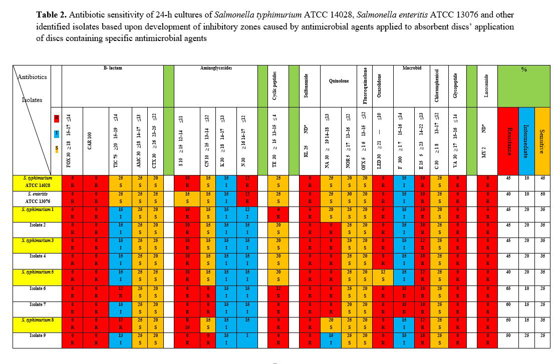
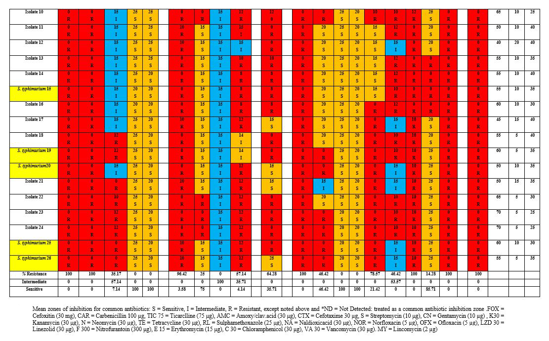
Antibiotic susceptibility profile for Salmonella spp.,
The results of susceptibility tests for S. typhimurium ATCC 14028 and S. enteritis ATCC 13076 were compared with results using Salmonella isolates. The bacterial isolates were obtained after overnight incubation following inoculation of single colonies into BHI media (Oxoid, U.K.). Cultures were spread on Mueller Hinton agar (Oxoid, U.K.), and individual plates were used for agar disk diffusion assays. Twenty different antibiotic-impregnated disks (Oxoid, U.K), with agents belonging to 7 different classes–b-lactam, aminoglycoside, cyclicpeptide, sulfonamide, quinolone, fluoroquinolone, and macrobid–were tested. Antibiotics were: cefoxitin (FOX, 30 μg, CT0119B), carbenicillin (CAR, 100 µg, CT0006B), ticarclline (TIC 75 μg, CT0167B), amoxicillin/clavulinic.acid (AMC, 30 µg, CT0223B), cefotaxime (CTX, 30 µg, CT0166B), streptomycin (S, 10 µg, CT0047B), gentamicin (CN, 10 µg, CT0024B), kanamycin (K, 30 μg, CT0025B), neomycin (N, 30 µg CT0033B), tetracycline (TE, 30 μg, CT0054B), sulphamethoxazole (RL, 25 µg CT0051B), naldioxic acid (NA, 30 μg, CT0031B), norfloxacin (NOR, 5 µg, CT0668B), ofloxacin (OFX, 5 µg, CT0446B), linezolid (LZD, 30 µg, CT1650B), nitrofurantoin (F, 300 µg, CT0036), erythromycin (E, 15 µg, CT0020B), chloramphenicol (C, 30 µg, CT0013B), vancomycin (VA, 30 µg, CT0058B) and lincomycin (MY, 2 µg, CT0027B). Diameters of zones of inhibition (mm) for each antibiotic and isolate were recorded using criteria recommended for Enterobacteriaceae [33]. Diameter measurements were used to classify isolates as sensitive (S) or resistant (R).