Breast cancer (BC) is one of the devastating malignancies and second-leading mortality cause in females worldwide. Traditional methods for BC screening include magnetic resonance imaging (MRI), ultrasound, mammography, or positron emission tomography (PET-scans). Unfortunately, these approaches possess several drawbacks such as high cost, low sensitivity, difficult operation with stressful conditions for patients [1, 2]. To date, several types of BC biomarkers such as nucleotides, cancer cells, proteins, and some small molecules have been explored to support a precise diagnosis. Of these biomarkers, the human epidermal growth factor receptor-2 (HER-2), a specific oncoprotein, is changed during the development and progression of BC. It was suggested the overexpression of HER-2 in breast cancer cells stimulates proliferation rate and tumor expansion [3, 4]. Currently, the diagnosis of HER-2+ BC is based on conventional techniques like cytological analyses using fine-needle aspiration (FNA), fluorescence in situ hybridization (FISH), gene expression monitoring, immunohistochemistry (IHC), and enzyme-linked immunosorbent assay (ELISA) procedures. Among the above-mentioned approaches, FNA and direct sampling are time-consuming and operationally invasive and can result in iatrogenic tumor implantation or metastasis [5]. Importantly, laboratory data have indicated that the level of HER-2 in the serum of BC patients ranged from 220 pM to 1.1 nM whereas these values reached 30 pM to 220 pM in healthy people, showing the much overlaps in the systemic levels of HER-2 under physiological and pathological conditions [6–8]. As a consequence, there is an urgent need to develop accurate and low-cost methodologies with the ability of rapid and sensitive recognition in BC patients.
Biosensors have been developed as key responses to circumvent the problems associated with HER-2 screening. Electrochemical biosensors are one of the most employed, highly sensitive, and easy to implemented tools for the detection of target molecules in biological samples [9–11]. One of the most appealing interests in electrochemical biosensing strategies is to use nano-biomaterials for the modification of electrode surface structure. The simultaneous application of nanomaterials in the electrochemical sensors can yield excellent achievements [12–14]. The synthesis protocol of nanomaterials is one of the limiting factors in their applicability, quality, final costs, and environmental viewpoints [15, 16]. Thanksgiving to colloidal procedures, electrosynthesis of nanomaterials is easily performed by dipping the working electrode in the electrochemical cell containing the precursor solution. A load of the same nanostructures on working electrodes using electrochemical methods is thought to increase detection outcomes [17–20]. In this case, there are no reducing, stabilizing agents in growth solution, which were applied in colloidal methodologies. Instead, a specified potential or current is implemented across the reaction cell to form nanoparticles. The integration of the electrosynthesis methods with the fast-growing nanotechnology field can present a unique gift to progress the synthesis chemistry for many applications [21–24].
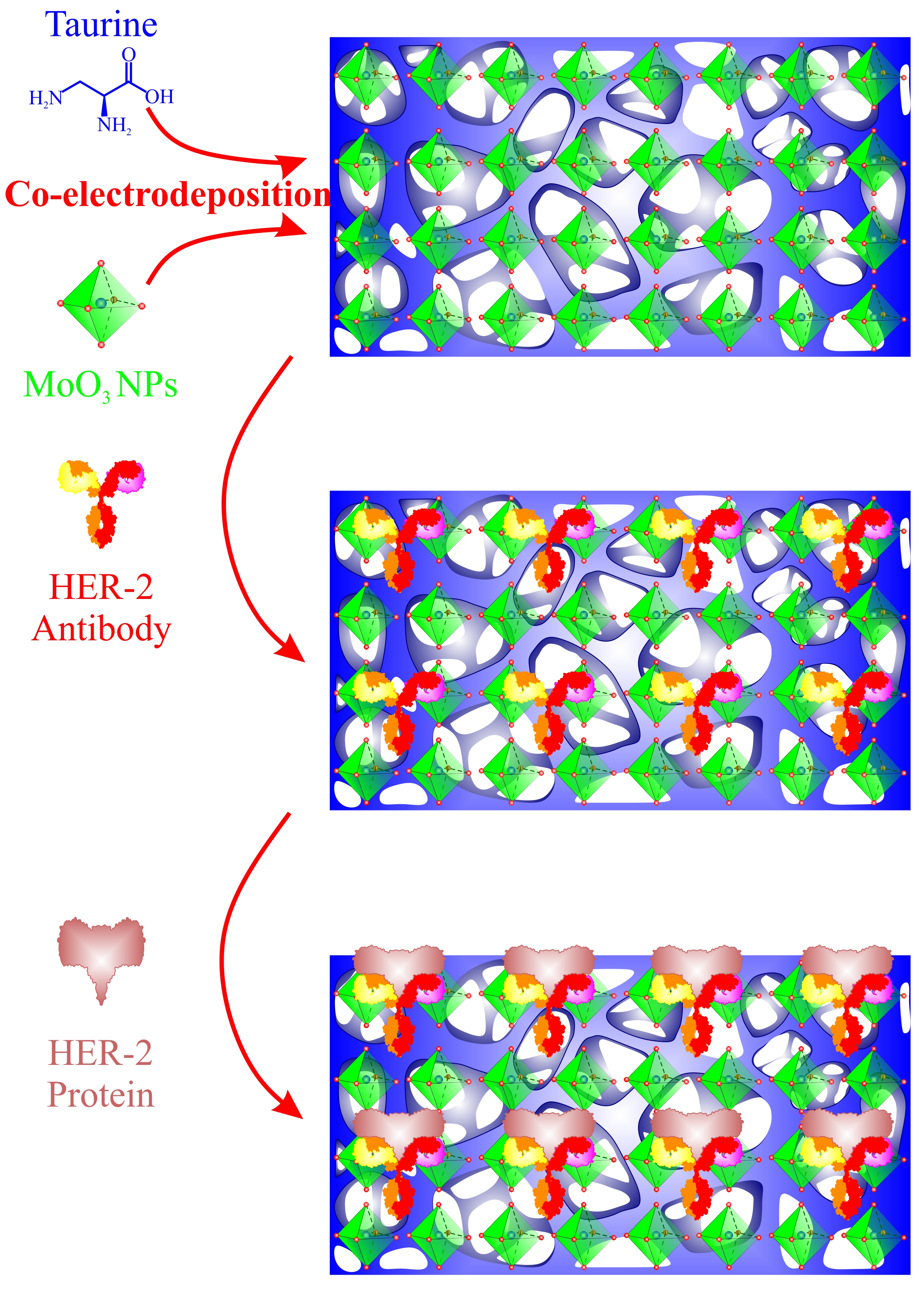
Molybdenum trioxide (MoO3) semi-conductor is of high interest among other semiconductors because of desirable biocompatibility, high electrical conductivity, big band gape, acceptable catalytic activity, tuning plasmon, large surface area, and lower final production costs [25, 26]. These superiorities facilitate the application of MoO3 in several domains such as electro-catalysis [27, 28], energy storage device development [29, 30], and fabrication of chemical sensors [31–34]. To this end, MoO3 nanomaterials have been used in different shapes, sizes, and functionalities in strategies associated with biosensing. One of the most prevalent interests in such an investigation is merging Mo nanostructures with other nanomaterials like platinum [35], graphene [36] and gold [31] nanomaterials. All these combinations are proposed to boost the performance of the Mo nanostructures and cover their application gaps like poor functionalibity and lower conductivity compared to the metal nanomaterials.
In this research, we used MoO3/poly-Tau (MoO3/p-Tau) nanofilms as a high-performance framework for detection of HER-2 in serum samples of BC patients. An electrosynthesis route was applied for the fabrication of the nanofilms onto the electrode surface. To the best of our knowledge, this is the first report of MoO3/p-Tau nano-films application in the bioassaying field. The highly homogeneous distribution of Mo nanoparticles into the p-Tau films was monitored using an appropriate potential range.