Preferential Enrichment of Inflammatory MiRNAs in CD11b + Cells from Brain and Bone Marrow
TaqMan RT-qPCR was used to measure selected inflammatory miRNAs including miR-146a-5p, miR-223-3p, miR-155-5p, and miR-150-5p levels in brain and bone marrow CD11b + cells from female and male naïve mice. While miR-146a and miR-150 levels were detected in bone marrow CD11b + cells in both females and males, they were more than 25-fold higher in brain CD11b + cells compared to bone marrow in both sexes (Fig. 1). Both miRNAs showed higher levels in brain CD11b + cells of male mice compared to females, but only miR-150 reached statistical significance. Conversely, miR-223-3p, and miR-155-5p were more than 25-fold higher in CD11b + cells isolated from bone marrow relative to brain in both sexes, with only miR-155-5p showing significant sex specific enrichment (males > females) in brain (Fig. 1). This pattern demonstrates that inflammatory miRNAs are differentially enriched in brain and bone marrow CD11b + cells.
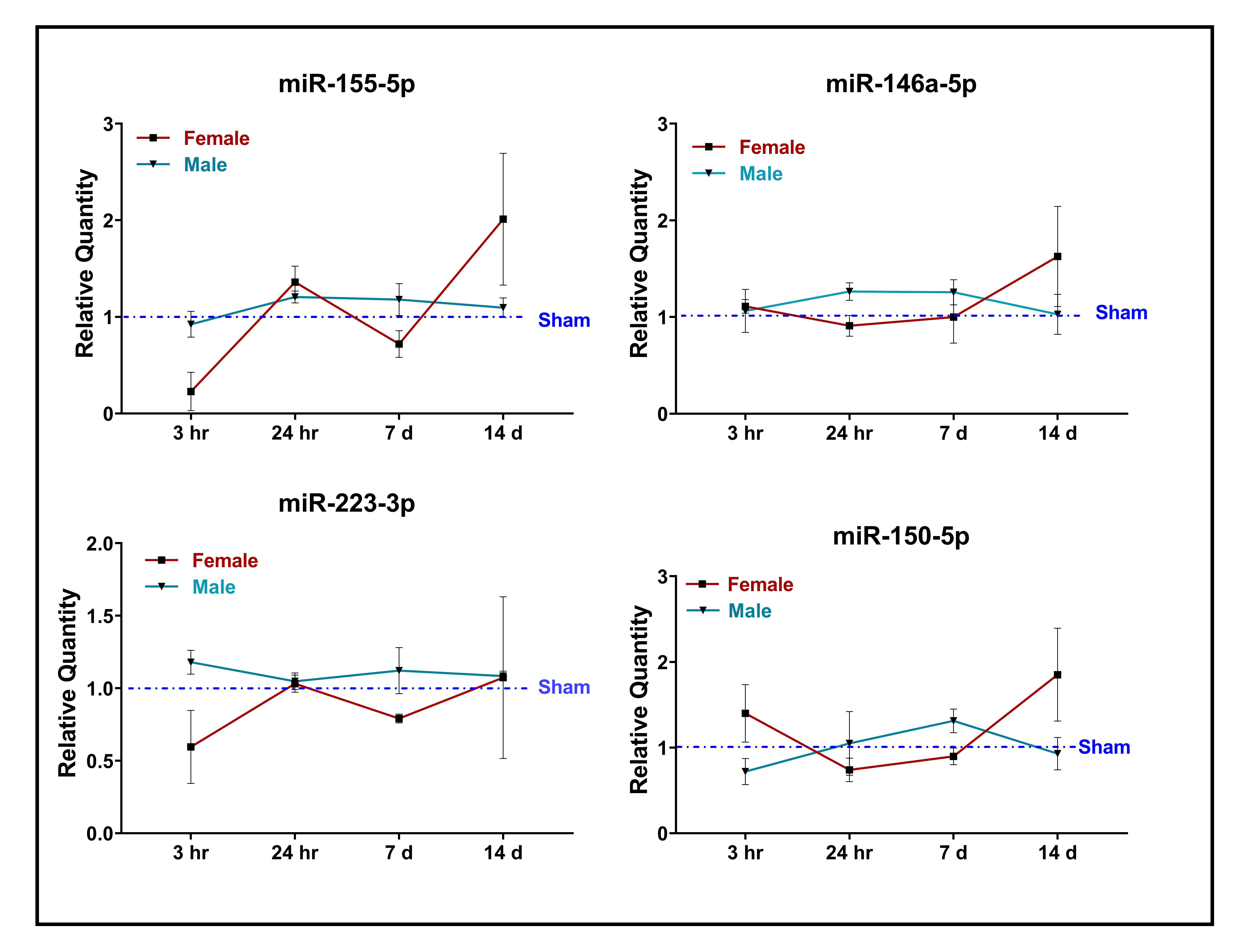
Sex-specific Alterations of Inflammatory MiRNAs in CD11b + Cells Following Severe TBI
To assess sex-specific miRNA alterations following TBI, we measured levels of these miRNA in CD11b + cells from injured cortical tissue and bone marrow at 3 hr, 24 hr, 7 days, and 14 days after severe TBI. Levels of pro-inflammatory miR-155-5p in brain CD11b + cells were significantly increased in both female and male mice after TBI, although females showed a significantly greater induction at 3 hr compared to males (Fig. 2a). The levels of anti-inflammatory miR-146a were altered marginally over time (Fig. 2b), while miR-223-3p levels significantly increased by 24 hr after TBI in both females and males, with females having significantly higher levels compared to males (Fig. 2c). MiR-150-5p was significantly increased in females but not males at 7 days and 14 days post-injury (Fig. 2d). Interestingly, we did not observe any TBI-related or sex differences in inflammatory miRNAs in bone marrow CD11b + cells (Suppl Fig. 1).
Sex-specific Alterations of Brain MiR-223-3p Targets and Inflammatory Gene Expressions in CD11b + Cells Following Severe TBI
We then examined the expression profile of miR-223-3p specific targets and related inflammatory genes in brain CD11b + cells of female and male mice following TBI. The sham animals did not exhibit any sex-specific patterns in the expression levels of any of the inflammatory genes examined. Reverse-correlated with the increase in miR-223-3p reported above (Fig. 2c), we found that the levels of two miR-223-3p targets, FBXW7 and TRAF6, significantly decreased at 24 hr post-injury only in brain CD11b + cells from female mice (Fig. 3a). Interestingly, we observed an increased expression of the anti-inflammatory gene, ARG1, in both female and male injured mice compare to sham animals at 24 hr post-injury, with females showing significantly higher expression than males (Fig. 3b). At this same 24 hr time point, brain CD11b + cells from female, but not male, mice had significantly higher expression of another anti-inflammatory gene, IL4 (Fig. 3b).
Several pro-inflammatory gene markers also exhibited a sex-related differential expression in brain CD11b + cells following TBI. For example, CCL2 was significantly increased in both female and male mice after TBI, although significantly greater increase was observed in males relative to females at 24 hr (Fig. 3c). In addition, there was a significant increase in COX2 expression only in male mice at 3 hr and 24 hr, which gradually decreased with time (Fig. 3c). The levels of TNFα and IL1b showed different sex-related expression patterns over time following injury (Fig. 3c). Specifically, both pro-inflammatory markers peaked at 3 hr after TBI in males and at 24 hr in females, with significantly higher levels in females compared to males at 24 hr (Fig. 3c).