Bacterial strains and routine culture of S. aureus
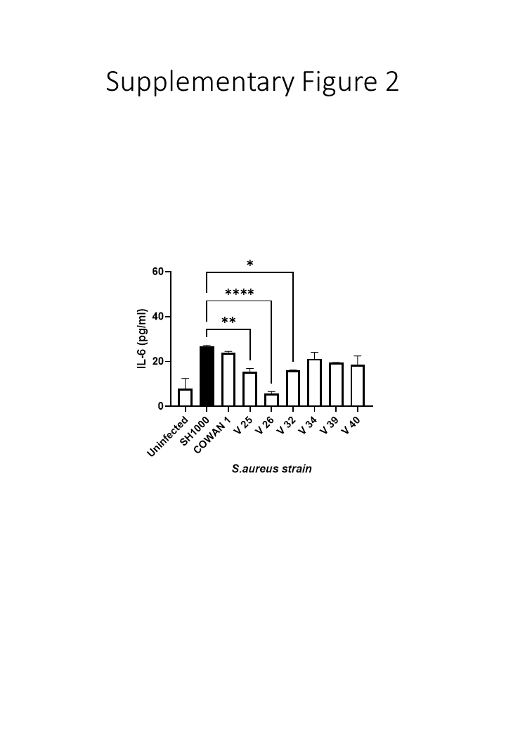
Six clinical methicillin resistant isolates obtained from human bronchoalveolar alveolar lavage fluid (BAL) were used [35, 78]. In addition, two control strains; S. aureus, SH1000 and S. aureus Cowan 1 ([79–84] and Table 2) were used. For culture, one single colony of S. aureus was taken from an agar plate and inoculated into 5ml of sterile tryptic soy broth (TSB), then grown overnight at 37°C. The overnight cultures were standardised to OD600 = 0.1 (~ 1 × 10⁸ cfu/ml).
Preparation and characterisation of Carbon Black (CB)
CB dose preparation
CB, AROSPERSE® 15 thermal black powder (Evonik Degussa GmbH #BT10506621) was weighed out at 1mg using the OHAUS Explorer Semi-Micro Balance housed in a WAYSAFE (#GP1540) to 1ml of the selected media (ultra-pure H2O, Tryptic soy broth (TSB) or 1% FBS/DMEM). This suspension was vortexed for 1 minute and sonicated in a 90W Ultrasonic water bath (Fisher Scientific #FB15046) at maximum power for approximately 30–40 minutes to ensure the CB was completely suspended. CB was then diluted to specific doses (including 0, 2, 4, 8, 10, 25, 50, 100 µg/ml) in water, TSB, 1% FBS / DMEM.
Zetasizer
Agglomerate medial and size distribution of CB samples was determined by dynamic light scattering (DLS) using a Malvern Zeta-Sizer Nano ZS (Malvern instruments Ltd., UK). Measurements were performed in deionised water, TSB and 1% FBS DMEM and presented as an average of 10 readings, with samples briefly vortexed and incubated at 37°C prior to measurements.
Transmission electron microscopy (TEM)
TEM was used to analyse CB particle size, shape, morphology, crystallinity and purity. A drop of diluted material (50 µg/ml in double distilled H2O) was drop-cast on a copper TEM grid coated with a continuous carbon film (Agar Scientific, UK) and left to air dry. TEM analysis was undertaken with a FEI Talos F200x G2 TEM (ThermoFisher Scientific, UK) operating at 200 kV and fitted with a high angle annular dark field (HAADF) detector, a Gatan Orius SC600A CCD camera, and an Oxford Instruments 80mm2 silicon drift energy-dispersive X-ray (EDX) spectrometer. Images were taken from 20 areas at magnifications between x7000 and x40000 with a dwell time of 10 µs.
S. aureus growth with CB
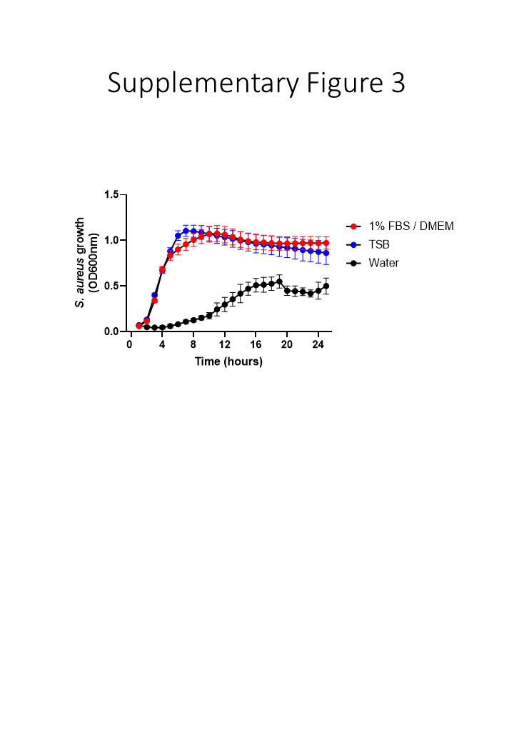
Standardised suspensions of S. aureus SH1000 were prepared as above but diluted to twice (2X) their final concentrations in water, TSB or 1%FBS/DMEM. Likewise, CB was also prepared at twice the concentration used (0, 4, 8, 16, 20, 50, 100 and 200µg/ml). Then using a 96 well plate, 100µL of diluted standardised S. aureus and 100µl of the double concentrated CB were combined (to generate the defined concentration) and left to incubate at 37°C at 200rpm for either 5 or 24 hours. Then the OD600 was read every hour, with constant oscillations (100 rpm), using the FLUOstar Omega microplate reader (BMG Labtech, Germany) for up to 24 hours. Preliminary experiments confirmed that CB at doses below 25µg/ml did not interfere with the measurement of absorbance (OD600nm) using the microplate reader (Supplementary Fig. 4)
Preparation of CB and S. aureus for SEM
Bacterial and CB suspensions were prepared as previously described above and then bound to Thermanox disks. Briefly, pre-sterilised Nunc™ Thermanox™ Coverslips (13mm diameter, Thermo Scientific, Paisley, UK) were taped onto a glass microscope slide and placed in a cytospin filter cartridge. Suspensions were mixed gently before 80 µl was pipetted into the cytospin cartridges prior to centrifugation at 112g for 3 minutes in a Shandon Cytospin 3. The cartridges were then taken apart and the Thermanox™ disk removed carefully. The samples were then left to air-dry overnight before being fixed with 2.5% glutaraldehyde in 0.1M piperazine-N,N′-bis(2-ethanesulfonic acid (PIPES), pH 7.4 for 5 min, post-fixed with 1% osmium tetroxide (OsO4; Simec Trade AG, Zofingen, Switzerland) in 0.1M PIPES (pH 6.8) for 1 h, dehydrated through an ethanol series (50%, 70%, 96%, 100%, 5 min each step) and an ethanol: hexamethyldisilazane (HMDS) series (2:1, 1:1, 1:2, for 5 min each step), and finally in 100% HMDS for 5min. Samples were then left to air-dry overnight, mounted on stubs and viewed with a Hitachi S4800 High resolution Scanning electron microscope using the upper secondary electron (SE) detector with a − 10 V or -20 V bias to minimise SE detection and maximise backscattered electron (BSE) detection, at an acceleration voltage of 1 kV and emission current of 10 µA. Images were taken from 10 random areas of the sample.
Cell culture
The choice of cells was determined by the major route of exposure for CB, namely skin and lung. Likewise, the lung and skin form the major sites for S. aureus pathogenesis. Therefore, epithelial cell lines from skin and lung were used in this study. HaCaT immortalized human keratinocytes, and A549 adenocarcinoma Type II human alveolar epithelial cells were used [85, 86]. Both cell lines were grown in Dulbecco’s, minimum essential medium (DMEM) supplemented with 10% fetal bovine serum (FBS), 1% penicillin (100µg/ml) / streptomycin (100U/ml), 1% glutamine (2mM) and incubated at 37°C / 5% CO2. HaCaT and A549s were sub-cultured at 90% confluence with TrypLE Express according to manufacturer’s instructions. Cell viability was assessed by trypan blue (0.2%) exclusion.
S. aureus infection of epithelial cells
A 10X standardised suspension of S. aureus SH1000 was prepared following overnight pre-culture (OD600 = 1.0, ~ 1 x10⁹/ml). Replicate 24 well plates were seeded with 50,000 epithelial cells / well in a total volume of 1ml cell culture media. The plates were then incubated at 37°C in a 5% CO2 environment for 24 hours. Then, the following day media was removed and replaced with 995µl of DMEM supplemented with 1% FBS and L-glutamine (without antibiotics). Then, 5 µl (~ 5 x 10⁶ bacteria) of 10X standardised S. aureus SH1000 were added before the plate was incubated for either 5 or 24 hours at 37°C in a 5% CO2 environment. Then the media was removed and decanted into a clean Eppendorf and centrifuged at 8064g for 5 mins. After centrifugation, 900µl of the supernatant was removed without dislodging the pellet and aliquoted into a clean Eppendorf before being placed in a -80 freezer for storage until ELISA determination could occur.
Co-exposure of epithelial cells to S. aureus and CB
Epithelial cell monolayers and S. aureus were prepared as previously described for infection studies with S. aureus SH1000 alone, except the following day media was removed and replaced with 495µl of DMEM supplemented with 1% FBS and L-glutamine (without antibiotics). Then, 5 µl (~ 5 x 10⁶ bacteria) of 10X standardised S. aureus SH1000 and 500 µl of CB dilutions (0-200µg/ml) were added before the plate was incubated for either 5 or 24 hours at 37°C in a 5% CO2. The media was then removed and decanted into a clean Eppendorf and centrifuged at 8064g for 5 mins. After centrifugation, 900µl of the supernatant was removed without dislodging the pellet and aliquoted into a clean Eppendorf before being placed in a -80 freezer for storage until ELISA determination could occur.
Enzyme linked immunosorbent assay (ELISA)
DuoSet ELISA (R&D Systems, Abingdon, UK) for human IL-8, IL-6 and IL-10 were carried out according to the manufacturers’ instructions. Human β-Defensin-2 (hβD-2) ELISA was assayed using a TMB development kit (PeproTech, London, UK) according to the manufacturer’s instructions.
Data and statistical analysis
Growth and cytokine data were presented as the mean ± standard error of the mean (SEM). A minimum of 3 biological repeats were conducted for all analyses presented. GraphPad Prism software (Version 9.1.2) was used for statistical analysis using a one-way ANOVA parametric test, including a Sidak post hoc test for multiple pairwise comparisons with *p ≤ 0.05 being considered significant.