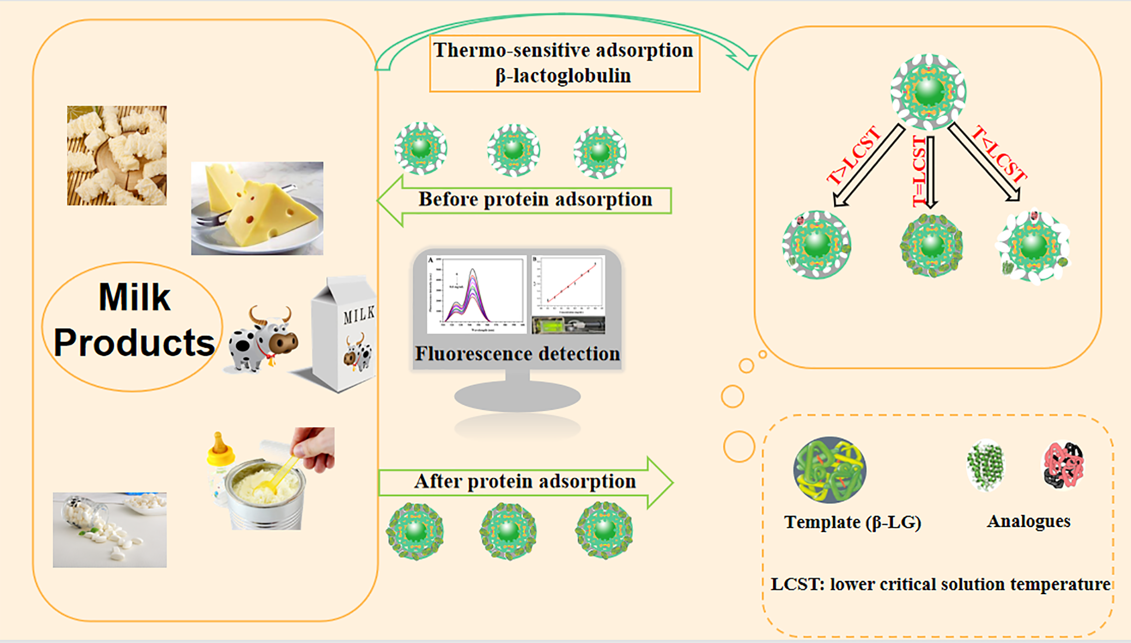
In this study, a thermo-sensitive molecularly imprinted fluorescence sensor was developed for the specific detection of β-Lactoglobulin (β-LG) allergen in milk products. The metal-organic frameworks (MIL-100) with a high specific surface area was coated on the surface of upconversion micro-particles (UCMPs). As the core, an imprinted polymer layer allowing for swelling and shrinking with response to temperature was prepared, which exhibited high adsorption and mass transfer capabilities for β-LG allergen. The fluorescence intensity of UCMPs@MIL-100@MIP decreased linearly with the concentration of β-LG in the range of 0.1–0.8 mg mL− 1 with detection limit of 0.043 mg mL− 1. The imprinting factor reached 3.415, which indicated excellent specificity of the UCMPs@MIL-100@MIP for β-LG allergen. In the analysis of β-LG allergen in actual milk samples, the proposed UCMPs@MIL-100@MIP fluorescence sensor produced reliable and accurate results (recovery: 86.0–98.4%, RSD: 2.8–6.8%), closely related to the results of standard HPLC method (correlation coefficient: 0.9949), indicating its feasibility in the detection of β-LG allergen.
Research
A UCMPs@MIL-100 based thermo-sensitive molecularly imprinted fluorescence sensor for effective detection of β-lactoglobulin allergen in milk products
https://doi.org/10.21203/rs.3.rs-915606/v1
This work is licensed under a CC BY 4.0 License
You are reading this latest preprint version
β-lactoglobulin
molecularly imprinted fluorescence sensor
upconversion micro-particles
metal-organic frameworks
thermo-sensitive
In recent years, the allergenicity of milk and its products has affected food safety, especially the healthy growth of infants and children, which has aroused widespread concern[1]. β-lactoglobulin (β-LG), belonging to the strong allergen lipocalin family, is one of the proteins that can cause strong allergic reactions[2]. According to the latest epidemiological survey results, approximately 2–7% of infants suffer from vomiting, diarrhea, rash and other food allergy symptoms due to the ingestion of β-LG, and about 82% of milk allergy patients are allergic to β-LG[3]. In the process of food processing, β-LG protein is also widely added to various foods to increase their nutrition, increasing the risk of exposure to β-LG in people with milk allergies[4]. In addition, milk products are also a special industry with great attention in the agriculture and animal husbandry industry chain. Therefore, the existence of β-LG in milk products will restrict the development of the milk industry and also have a certain negative impact on the development of agriculture and animal husbandry food. Therefore, it is necessary to develop a convenient, accurate, sensitive and specific analytical method to monitor the content of β-LG in milk products, which is not only convenient for manufacturers to accurately label the allergen on food packaging, but also beneficial to protect consumers from the threat of allergies. Currently, various analytical strategies based on different principles have been developed for the quantitative analysis of β-LG in milk products, such as high-performance liquid chromatography (HPLC) method, sandwich ELISA, surface plasmon resonance method, electrochemical method[5–8]. These methods have remarkable advantages in terms of detection accuracy, sensitivity and reproducibility, but often require expensive equipment or environmentally sensitive recognition molecules (antibodies, etc.), which not only increase the cost and time of detection, but also are not conducive to on-site, real-time or popular detection. Fluorescence sensing technology has received extensive attention in the field of food safety detection because of its high sensitivity, fast response speed, simple operation, easy integration and miniaturization. Up to now, the use of fluorescence sensing in the detection of β-LG allergen in milk products has rarely been reported. Therefore, it is feasible and significant to design an effective fluorescence sensor for β-LG in milk products.
Upconversion micro-particles (UCMPs) are common fluorescent materials doped with lanthanide rare earth elements[9]. Under the excitation of low-energy infrared or near-infrared light, UCMPs can emit high-energy visible light due to its unique anti-coking luminescence characteristics[10]. Compared with ordinary quantum dots and organic fluorescent dyes, UCMPS has the advantages of low toxicity, good biocompatibility and low biological fluorescence background value, which makes its application in detection, biomarkers and in vivo imaging in complex systems increasingly attracted attention[11–13]. In comparison with single component materials, composite materials with a core-shell structure can be customized according to actual needs, and are easily modified, showing better performance. Metal-organic framework materials (MOFs) are typical shell materials with large specific surface area, uniform pore size distribution and adjustable pore structure. These properties make MOFs materials very promising in the fields of gas storage, catalysis, separation, drug transportation and photoelectric sensing[14–18]. MIL-100 (Fe) is one MOFs material composed of central particles Fe and ligands trimesic acid (H3BTC), which has a large specific surface area and good thermal stability. Several composites using MIL-100 shell, such as Fe3O4/MIL-100, Au/MIL-100, have been developed for enrichment and detection of various targets[19, 20]. However, there are few reports on combining MIL-100 (Fe) and UCMPs to construct core-shell composites with biomacromolecule recognition capabilities.
Molecularly imprinted polymers (MIPs), which contain abundant imprinted sites that match the size, shape, and functional groups of template molecules, are defined as artificial antibodies[21]. MIPs is an effective substitute for natural antibodies due to its simple preparation and high stability[22, 23]. Up to now, MIPs with good specificity and selectivity to various small molecules and proteins have been developed and applied to the target detection and enrichment in different samples[24, 25]. However, traditional MIPs have defects such as limited mass transfer, structural rigidity, poor accessibility to target molecules and uncontrollable adsorption process, leading to a decrease in the adsorption capacity, rate and selectivity of MIPs for analytes[26]. Stimulus-responsive MIPs are known as environmental responsive MIPs because of their phase transformation and structural transformation characteristics in response to external stimuli such as temperature, pH, magnetism and light[27–29]. Among them, the thermo-sensitive MIPs have remarkable adsorption reversibility and stability, which attracted widespread attention[30]. The MIPs prepared using N-isopropylacrylamide (NIPAM) as the monomer have a flexible structure and can exhibit a reversible expansion and contraction transition according to temperature changes[31]. According to the previous reports on thermo-sensitive MIPs, such "on demand" materials have shown great potential in the field of biomolecular recognition, but the adsorption and recognition capabilities remain to be improved.
Based on the above considerations, this study proposed a feasible and effective strategy for the detection of β-LG in milk products. A core-shell composite with UCMPs as core and MIL-100 (Fe) as shell was prepared and used as a carrier to develop fluorescent thermo-sensitive MIP materials. This material combined the fluorescence characteristics of UCMPs, the high specific surface area of MIL-100 (Fe) and the high specificity recognition of thermo-sensitive MIP, and has a specific, rapid and stable fluorescence response to β-LG, which can be used for convenient and efficient analysis of β-LG allergen in milk and its products.
Materials and chemicals.
The proteins β-LG (pI 5.1–5.3, 90%), α-lactalbumin (ALa, pI 4.2–4.5, 85%), lactoferrin (Lf, pI 8.0, 95%), and casein (Cas, pI 4.8, > 95%) were obtained from Yuanye Biotech Co. Ltd. (Nanjing, China). Y(CH3COO)3·H2O (99.9%), Yb(CH3COO)3·4H2O (99.9%), Er(CH3COO)3·4H2O (99.9%), N,N-methylenebisacrylamide (MBA), oleic acid (OA, 90%), ethylene glycol (EG, > 99%), N,N,N',N'-tetramethylethylenediamine (TEMED, 99%), polyacrylic acid (PAA), and NIPAM, were provided by Macklin Biotech Co. Ltd. (Shanghai, China). Trisodium citrate dihydrate (TSC, 99.0%), ammonium persulfate (APS), NaCl (99.5%), NH4F, FeCl3·6H2O, and H3BTC were purchased from Aladdin Reagent Co., Ltd. (Shanghai, China). The reagents anhydrous ethanol, acetic acid, trifluoroacetic acid (TFA, > 99.9%), and acetonitrile at least of analytical grade were acquired from National Medicine Group Chemical Reagent Co., Ltd. (Shanghai, China). Deionized water (18.2 MΩ cm) was obtained by a Water Pro water purification system (Labconco, Kansas City, USA).
Instrumentation.
The UV absorbance at a wavelength of 280 nm was recorded on the Evolution 300 UV-Vis Spectrophotometer (Thermo, USA). Infrared spectra were obtained using a Fourier transform infrared spectrometer (Nicolet iS 50, Thermo, USA). Fluorescence measurements were performed on an F-7100 fluorescence spectrophotometer (Techcomp, Shanghai, China) equipped with a 980 nm external exciter (2 W, Shanghai Feibo Laser Technology Co., China) as the light source. Transmission and scanning electron microscopy (TEM and SEM) images were obtained from a Talos G2 200X and Apreo electronic microscope (Thermo, USA), respectively. X-ray powder diffraction (XRD) patterns were recorded on a Malvern Panalytical apparatus at a scanning rate of 1° min− 1 in the 2θ range from 5° to 80° (Shanghai, China). Energy dispersive X-ray photoelectron spectroscopy (XPS) patterns were measured on ARL QUANT'X Energy dispersive X-ray spectrometer (Thermo, USA). Nitrogen adsorption/desorption analysis was performed on a 3H-2000PS apparatus (Beijing, China) with a bath temperature of 77 K. Millipore ultrafine filters from Merck Company (Germany) and a centrifuge machine from Eppendorf (Germany) were used in the pre-treatment of milk products. A HPLC system equipped an SPD-20A detector from Shimadzu Corporation (Japan) was applied to verify the results of β-LG measurement in milk products.
Synthesis and Modification of UCMPs.
Hexagonal NaYF4: Yb3+, Er3+ UCMPs were synthesized refer to the previous work with a little modification[32]. At room temperature, 8 mmol of TSC were dissolved in 20.0 mL of H2O in a 100.0 mL reaction flask. Another 10.0 mL of homogenous aqueous solution containing 1 mmol of rare earth salt (Y3+ : Yb3+ : Er3+ = 0.78 : 0.2 : 0.02) was added and the mixture was stirred magnetically for 10 min. Then, 10.0 mL of a mixed solution containing 337.0 mg (5.76 mmol) of NaCl and 444.0 mg (12 mmol) of NH4F was added into the flask dropwise, gradually forming a milky solution. Subsequently, 20.0 mL of OA and 10.0 mL of EG were slowly added into the milky solution successively. After stirring for another 1.5 h, the mixture was transferred into a 100.0 mL stainless-steel autoclave for solvothermal reaction in Muffle furnace, sealed and kept at 180°C for 6 h. After the autoclave was cooled to room temperature, the white products (UCMPs-OA) were obtained by centrifugation, fully washed with ethanol and dried at 60°C.
Furthermore, an exchanging process of OA and PAA ligand was carried out to ensure that the fluorescence probe UCMPs could be applied effectively for protein detection. 100.0 mg of UCMPs-OA product were washed thoroughly with ultrasound in 10.0 mL of ethanol (adjusted with HCl) at pH 1.0, and then dispersed in 10 mL of ethanol solution after centrifugation. The dispersion was dropped into ethanol solution containing 30.0 mg of PAA and stirred magnetically at room temperature for 12 h. After removing excess PAA by centrifugation and ethanol washing, the product UCMPs-PAA was dried at 60 ℃ for 10 h.
Synthesis of UCMPs@MIL-100.
The detailed synthesis process of UCMPs@MIL-100 was as follows. 100.0 mg of UCMPs-PAA product was mixed with 30.0 mL of ethanol in a reaction vessel thoroughly. 13.5 mg (0.05 mmol) of FeCl3·6H2O was added into the vessel and the mixture was magnetically stirred for 30 min at room temperature to allow a stable connection between Fe3+ and UCMPs-PAA. Subsequently, 10.0 mL of an ethanol solution containing 10.5 mg (0.05 mmol) of H3BTC was added and the mixture was stirred and heated in a water bath at 40 ℃ for 40 min, and the solid product was collected by centrifugation. The same procedure was repeated 10 times to coat MIL-100 uniformly on the surface of UCMPs. The final product was washed alternately with water and ethanol and dried at 60 ℃ for 10 h in a vacuum drying oven to obtain UCMPs@MIL-100 light yellow powder.
Synthesis of UCMPs@MIL-100@MIP and UCMPs@MIL-100@NIP.
Accurately weighted β-LG (10.0 mg), and the prepared UCMPs@MIL-100 (50.0 mg) were dispersed in 10.0 mL of water in a 25.0 mL round-bottom flask and shaken for 10 min. Subsequently, 79.0 mg (0.7 mmol) of NIPAM and 162.0 mg (1.05 mmol) of MBA were added, and then shaken at room temperature for 1 hour. After adding 10.0 mg of APS and 100.0 µL of TEMED (5%, v/v) and nitrogen bubbling for 10 min to remove oxygen, the polymerization reaction was carried out at 32°C for 24 h. After the polymerization was completed, the template β-LG was alternately eluted with 0.5 mol L− 1 NaCl and acetic acid (6.0 vt%) at 20 ℃ for several times, and until no β-LG was detected by UV. The UCMPs@MIL-100@NIP was prepared adopting the same procedure except the addition of β-LG.
Fluorescence Measurement and Thermo-Sensitive Adsorption Experiments.
All fluorescence analysis were performed using an F-7100 fluorescence spectrometer equipped with with a 980 nm external exciter, which recorded emission wavelengths in the 400–700 nm range. 2.0 mg of UCMPs@MIL-100@MIP was mixed with 2.0 mL of β-LG solution with a certain concentration in a 5.0 mL centrifuge tube. After being shaken for 1 h, the mixture was measured the fluorescence intensity (λ = 544 nm) quickly.
The adsorption performance of UCMPs@MIL-100@MIP and NIP at different temperature was investigated by mixing 2.0 mg of the prepared UCMPs@MIL-100@MIP or NIP with 2.0 mL of β-LG solution (0.2 mg mL− 1) and shaken at different temperatures (20, 32, 44 ℃) for 1 h. After centrifugation at 5000 rpm, the concentration of β-LG in supernatant was measured by UV-vis spectrophotometer.
Sample Preparation and Validation of Method.
In this study, raw milk and infant formula were selected as actual samples to study the application capability of the prepared UCMPs@MIL-100@MIP fluorescence sensor. Accurately weighted raw milk (5.0 mL) or infant formula (2.0 g), were placed into a 25.0 mL volumetric flask, respectively. After reaching a constant volume and adjusting pH to 4.6 using acetic acid, the mixture was allowed to stand for 1 h and centrifugated for 10 min (5000 rpm). The collected supernatant was filtered through a 0.22 µm filter, immediately afterwards, the raw milk supernatant was diluted 2 times, and analyzed using the developed fluorescence sensor. The resulting fluorescence intensity was used to calculate the amount of β-LG. The HPLC separation was achieved on a Hypersil GOLD C8 column (4.6 × 250 mm, 3.5 µm) using the mixture of H2O (0.1% TFA) (A) and acetonitrile (0.1% TFA) (B) as the mobile phase. The gradient conditions at a flow rate of 1.0 mL min− 1 were as follows: A, from 70–50% (0–15 min); A, from 50–70% (15–17 min). The detection wavelength and injection volume were 280 nm and 10.0 µL, respectively. The results of β-LG analysis from the proposed fluorescence sensor and HPLC were compared, and the correlation coefficient (R2) was calculated.
Preparation of UCMPs@MIL-100@MIP
This study integrates the fluorescence characteristics of UCMPs, the high specific surface area of MIL-100 material, and the specific recognition capability of thermo-sensitive MIP to develop a fluorescence strategy for the specific detection of β-LG. Scheme 1A shows the synthesis process of NaYF4: Yb3+, Er3+ UCMPs with green fluorescence by solvothermal method. Through the ligand exchanging process between OA and PAA, the prepared UCMPs were transformed into hydrophilic UCMPs (Scheme 1B). Because of the interaction between Fe3+ and the carboxyl groups of UCMPs and H3BTC, MIL-100 framework was wrapped around the hydrophilic UCMPs. The template protein β-LG can be fixed onto the surface of UCMPs@MIL-100 by non-covalent interaction, and the MIP layer was prepared by polymerizing NIPAM and MBA in aqueous solution. After elution of template protein β-LG, UCMPs@MIL-100@MIP with specific recognition sites was obtained.
Due to the use of thermo-sensitive monomer NIPAM, the adsorption and desorption performance of UCMPs@MIL-100@MIP can be achieved by controlling the external temperature. When the temperature is lower than lower critical solution temperature (LCST), NIPAM exhibits hydrophilicity, and the expanded imprinting cavities have no complementary affinity to the template protein functionally and spatially. When the temperature is higher than the LCST, the NIPAM is in a hydrophobic state, and the imprinting cavity shrinks. At this time, although the imprinting cavity is not complementary to the target protein, the hydrophobic interaction plays a leading role and the protein will also be captured.
Characterization of UCMPs@MIL-100@MIP
SEM and TEM analysis. SEM and TEM were used to observe the surface morphology and size of the synthesized UCMPs, UCMPs@MIL-100 and UCMPs@MIL-100@MIP (Fig. 1). Evidently, the bare UCMPs showed regular hexagonal with a particle size of about 1.5 μm and a thickness of about 188 nm (Fig. 1A,B). When MIL-100 film was formed on the surface, the size of the UCMPs@MIL-100 increased significantly and the coating thickness of MIL-100 film was about 80 nm (Fig. 1C,D). Accompanied by the growth of MIP upon the surface of UCMPs@MIL-100, the size of the coating layer has expanded to 162 - 188 nm (Fig. 1E,F), and the shape was irregular, confirming the successful preparation of UCMPs@MIL-100@MIP.
FT-IR spectra analysis. The UCMPs-PAA, UCMPs@MIL-100 and H3BTC were characterized by FT-IR spectroscopy. At 2850 and 2918 cm-1, there were symmetrical stretching vibration peaks and anti-symmetric stretching vibration peaks, respectively, which suggested the stretching vibration of the -CH2- on PAA (Fig. 2A (a)). The characteristic peaks at 1421 and 1636 cm-1 were attributed to the stretching vibration of -COOH groups. These results have demonstrated that the UCMPs with PAA ligand were successfully synthesized.
The characteristic peaks at 2664, 1720 and 918 cm-1 in Fig. 2A(c) correspond to the stretching vibration of the O-H, C=O, and the bending vibration of the O-H in H3BTC, respectively. In the FT-IR spectra of UCMPs@MIL-100 (Fig. 2A(b)), these three main characteristic peaks disappeared, and two significant absorption peaks appeared at 1622 and 1379 cm-1, which related to the symmetric and asymmetric stretching vibrations of ionized -COO-, respectively. This indicated that the -COOH groups of H3BTC dissociated into -COO- anions and formed coordination bonds with Fe3+. In addition, the fingerprint peaks derived from the vibration of the benzene ring were observed at 760 and 712 cm-1. These FT-IR spectra results were in full agreement with the step-by-step assembly process of MIL-100, indicating the surface of UCMPs has been successfully coated by MIL-100 framework.
XRD and XPS analysis. Fig. 2B has illustrated the crystal phase structure of UCMPs, UCMPs@MIL-100 and UCMPs@MIL-100@MIP determined by powder Xray diffraction. In the powder XRD diagram of UCMPS@MIL-100, UCMPS diffraction peaks were observed to be well preserved, and part of the characteristic peaks were consistent with those of MIL-100 previously reported, suggesting that the composite material was composed of UCMPS and MIL-100. In addition, the peak intensity of UCMPS@MIL-100@MIP was significantly reduced compared with that UCMPS@MIL-100. The peak intensity reflected the crystallization of the material, so it is speculated that the reason for the weakening was the formation of the MIP film on the surface. These results confirmed the successful prepare of UCMPS@MIL-100@MIP, which was consistent with the results of above characterization results.
To further demonstrate the component of UCMPs@MIL-100 and UCMPs@MIL-100@MIP, the composites obtained were characterized using XPS. In Fig. 2C, the primary signals of C1s at 284.81 and 288.6 eV, O1s at 531.71 eV and Fe2p at 712.09 eV can be clearly observed, illustrating the MIL-100 was successfully coated on the surface of the UCMPs. Compared with Fig. 2C, the N1s peak was clearly observed at 397.02 eV in Fig. 2D. The N source mainly comes from the N elements carried by β-LG, NIPAM, and MBA during the formation of the MIP layer, indicating the MIP was successfully coated on the surface of UCMPs@MIL-100.
Fluorescence quenching mechanism of β-LG to UCMPs@MIL-100@MIP.
In the study, the prepared UCMPs and UCMPs@MIL-100 materials (1.0 mg) were dispersed in 2.0 mL of water to investigate the fluorescence properties. As shown in (Additional file 1: Fig. S1A), under the excitation of the external 980 nm laser, UCMPs and UCMPs@MIL-100 appear green fluorescence emission peaks at 529 nm and 544 nm, respectively, which is due to the transition of Er3+ between the 2H11/2→4I15/2 and 4S3/2→4I15/2 energy levels. Therefore, the maximum emission peak at 544 nm was selected as a marker to evaluate the fluorescence characteristics of the synthesized materials. The fluorescence intensity of UCMPs@MIL-100 was significantly lower than that of UCMPs, which is due to fluorescence quenching of UCMPs caused by MIL-100 coating. These results preliminarily prove the successful synthesis of UCMPs@MIL-100 composites.
As shown in (Additional file 1: Fig. S1B), compared with UCMPs@MIL-100@NIP (a), UCMPs@MIL-100@MIP without removing β-LG has lower fluorescence intensity (c). While β-LG was removed, the fluorescence intensity of UCMPs@MIL-100@MIP (b) was significantly enhanced, almost close to that of NIP, which verified the quenching effect of β-LG on the fluorescence of UCMPs. Current studies have proved that the main mechanisms that cause fluorescence quenching are fluorescence resonance energy transfer (FRET) and photoinduced electron transfer (PET). Nevertheless, FRET occurs when the excitation band of the fluorescent acceptor and the emission band of the donor overlap in the analysis system. According to (Additional file 1: Fig. S1C), the absorption peak of β-LG at 280 nm did not overlap with the emission peak of the fluorophore. Therefore, the fluorescence quenching effect of β-LG on UCMPs is probably caused by electron transfer.
Thermo-sensitive property of the UCMPs@MIL-100@MIP.
As is known to all that NIPAM-based polymers exhibit both hydrophilic and hydrophobic state at different temperatures, simultaneously, the volume of polymers would change with the external temperature. As a result, the influence of temperature on the adsorption capacity of the prepared UCMPs@MIL-100@MIP was investigated. (Additional file 1: Fig. S2) showed the fluorescence intensity of the UCMPs@MIL-100@MIP without adding the template protein β-LG at 20 °C and 44 °C. Fig. 3A showed the fluorescence intensity of the UCMPs@MIL-100@MIP in adsorption to β-LG at 20 °C and 44 °C, indicating a significant temperature dependence of their interactions. After five cycles, the fluorescence intensity of the thermo-sensitive UCMPs@MIL-100@MIP was almost unchanged, indicating its good fluorescence anti-attenuation ability. These results demonstrated that β-LG adsorption and desorption of UCMPs@MIL-100@MIP can be realized by controlling the external temperature, which provides the basis for its repeatable use in fluorescence sensing.
Furthermore, the adsorption capacity (Q, mg g-1) of UCMPs@MIL-100@MIP and NIP for β-LG was calculated by the following equation.

In which, C0 and C represents the initial and residual concentration of β-LG, mg mL-1, respectively; V is the volume of β-LG solution, mL; and m represents the mass of MIP or NIP, g.
As shown in Fig. 3B, the adsorption capacity of the prepared UCMPs@MIL-100@MIP for β-LG reached 183.0 mg g-1 at 32 °C, which was significantly higher than that at 20 °C (47.0 mg g-1) and 44 °C (90.9 mg g-1). This is because at the temperature, the shape and size of the imprinted sites or cavies formed in the polymer were complementary to β-LG. At 20 °C, the NIPAM monomer is hydrophilic and forms a large number of hydrogen bonds in water, which enlarged the imprinting cavity of the polymer and resulted in most β-LG molecules entering and leaving unrecognized. At high temperature (44 °C), the hydrogen bonds formed by NIPAM were destroyed and the hydrophobic action dominated, leading to shrinkage of the polymer cavity in the aqueous phase. This hydrophobic effect also leads to a significant increase in non-specific adsorption, making its adsorption capacity higher than 20 °C, which is consistent with the study of Zhou et al[33]. In addition, UCMPs@MIL-100 material had a larger specific surface area than traditional carrier materials, reaching 637.38 m2 g-1, measured by nitrogen adsorption/desorption isotherm. This was also one important reason why UCMPs@MIL-100@MIP had stronger adsorption performance for the target protein.
Optimization of UCMPs@MIL-100@MIP preparation conditions.
In the study, the amount of UCMPs@MIL-100, the molar ratio of functional monomer and cross-linker, and the adsorption environment (pH) were investigated to obtain the optimal adsorption performance of UCMPs@MIL-100@MIP. The variable control method was adopted, and the imprinting factor (IF) was used as the final judgment result. The amount of UCMPS@MIL-100 prepared as the fluorescence source is closely related to the sensitivity of the constructed fluorescence sensor. (Additional file 1: Fig. S3A) showed the fluorescence responses of MIP and NIP prepared using different amounts of UCMPS@MIL-100. It can be observed that the addition amount of UCMPS@MIL-100 significantly affects the fluorescence response of the prepared MIP and NIP. When the addition amount is 50 mg, the maximum value of IF is 2.465. The cross-linker can form a stable rigid structure, which is conducive to the curing of the functional monomer in the polymerization layer, and then forming cavities or binding sites that match the template molecules. When the cross-linker is insufficient, the network structure of imprinting layer cannot be well connected, which affects the adsorption of β-LG molecule by MIP. Nevertheless, superfluous cross-linker will increase the thickness of the imprinted layer, resulting in mass transfer barrier, which will not only affect the mass transfer speed of β-LG in the imprinted layer, but also hinder its interaction with the fluorescence source UCMPS@MIL-100, thus reducing the detection sensitivity of the fluorescence sensor. By comparing the fluorescence response of UCMPs@MIL-100@MIP and NIP prepared under different ratios of functional monomers and cross-linker (Additional file 1: Fig. S3B), the IF reaches the maximum value (2.790) at the molar ratio of 2/3, which was chosen for further experiments. In addition, when the adsorption environment pH is 7.4 (Additional file 1: Fig. S3C), the best IF value of 3.208 is obtained. This is because when the pH of the solution is lower than 7.4, the positive charge on the surface of β-LG is less, while the alkalinity of UCMPs@MIL-100@MIP and the solution environment is relatively weak. When pH = 7.4, the surface positive charge of β-LG increases, and the alkalinity of UCMPs@MIL-100@MIP was stronger than that of solution system, indicating that UCMPs@MIL-100@MIP plays an important role in the recognition and retention of β-LG. With the continuous increase of pH value, the affinity of the solution system to β-LG gradually dominates, leading to gradually lost the recognition ability of UCMPS@MIL-100@MIP.
Fluorescence response of UCMPs@MIL-100@MIP to β-LG.
In this work, the fluorescence response of prepared UCMPs@MIL-100@MIP and NIP to different concentrations of β-LG allergen was evaluated. As shown in Fig. 4C, the fluorescence response value (F0/F) of UCMPs@MIL-100@MIP is significantly correlated with the concentration of β-LG in the range of 0.1 - 0.8 mg mL-1, in line with the following Stern-Volmer equation.

In which, F0 and F respectively represent the fluorescence intensity before and after the adsorption of β-LG, KSV is the quenching constant, and C represents the β-LG concentration (mg mL-1).
The fluorescence quenching equation of UCMPs@MIL-100@MIP is F0/F = 1.4423 C + 0.9538 with R2 of 0.9881, and the LOD was calculated as 0.043 mg mL-1. Compared with the fluorescence spectra of NIP at the same β-LG concentration, the quenching degree of UCMPs@MIL-100@MIP is obviously higher than that of NIP (Fig. 4A and 4B). This is because more binding cavies or recognition sites matching the size and shape of β-LG protein are formed in the imprinting layer. By comparing the slope of the fluorescence quenching equation (Fig. 4C), the IF was calculated as 3.415, indicating that the prepared UCMPs@MIL-100@MIP had good selectivity and specificity for β-LG recognition.
Kinetics evaluation of UCMPs@MIL-100@MIP.
To evaluate the kinetic properties of the prepared UCMPs@MIL-100@MIP and NIP, the equilibrium binding analysis was performed at a β-LG concentration of 0.4 mg mL-1. As can be seen from (Additional file 1: Fig. S4), the adsorption rate of UCMPs@MIL-100@MIP increases within 30 min and almost reached the adsorption equilibrium within 60 min. In the same period of adsorption, the F0/F change of UCMPs@MIL-100@MIP for β-LG was more significant than that of UCMPs@MIL-100@NIP. This is because UCMPs@MIL-100@MIP generates imprinting sites with respect to β-LG during the preparation process and has specific and non-specific binding during the adsorption process. However, UCMPs@MIL-100@NIP only existed non-specific adsorption. In addition, these results also indicated that the introduction of MIL-100 material not only increased the number of β-LG specific recognition sites in imprinting system, but also arranged the specific recognition sites in order, which was beneficial to the rapid binding of β-LG. This verifies the merits of this work in improving the adsorption capacity and efficiency of the molecularly imprinted system.
Selectivity Study.
The selectivity of UCMPs@MIL-100@MIP was evaluated using ALa, Lf, and Cas as competitive proteins at 0.4 mg mL-1 concentration. As illustrated in Fig. 5A, it was clearly observed that the F0/F of UCMPs@MIL-100@MIP for β-LG changes more significantly than ALa, Lf, and Cas. However, there was no significant difference in F0/F of the selected proteins for UCMPs@MIL-100@NIP. The IF was calculated as 2.19, 1.27, 1.21, and 1.15, respectively. This is because the specific cavies or recognition sites are formed that complement the size, shape, and functional groups of β-LG protein during the preparation of UCMPs@MIL-100@MIP. However, due to the lack of template protein β-LG, UCMPs@MIL-100@NIP only forms non-specific adsorption sites, resulting in the fluorescence of UCMPs not being significantly quenched by the target protein.
Equal amounts of the interfering proteins were added into the β-LG solution (0.4 mg mL-1) to further investigate the anti-interference ability of the developed UCMPs@MIL-100@MIP. As shown in Fig. 5B, the fluorescence response of UCMPs@MIL-100@MIP showed no significant changes in the three-protein mixed system compared to β-LG, indicating that its specific recognition ability for β-LG was not affected by the interfering proteins. UCMPs@MIL-100@NIP obtained a higher fluorescence response in a mixed protein system than each single interfering protein. These results indicate that the prepared UCMPs@MIL-100@MIP has significant specificity for β-LG recognition and can be applied under the hindrance of the interferents in complex samples.
Sample analysis and method validation.
To evaluate the application capability of the developed fluorescence sensor for analyzing β-LG in actual samples, raw milk and infant formula were selected and spiked with β-LG at three levels (0.1, 0.2, and 0.4 mg mL-1). After simple sample treatment, the β-LG content of the resulting extracts was measured using the molecularly imprinted fluorescence sensor and validated by standard HPLC method. Table 1 illustrates the measurement results of β-LG content obtained. The data listed is from a 10-fold dilution of raw milk extract and a 5-fold dilution of infant formula milk powder.
Obviously, at all concentrations tested, the proposed fluorescence sensor yielded β-LG content similar to those obtained using HPLC, with a correlation coefficient achieving 0.9949 (Additional file 1: Fig. S5). This means that the fluorescence sensor prepared in this study can be used for reliable and accurate analysis of β-LG. A comparison of the results of the reported strategies for β-LG analysis in various matrices was provided in Table 2, highlighting the merits of the developed UCMPs@MIL-100@MIP fluorescence sensor.
In this study, the fluorescence characteristics of UCMPs, the high specific surface area of MIL-100 and the high selectivity of molecular imprinting technology were effectively combined to prepare a thermo-sensitive molecular imprinted fluorescence sensor for the detection of β-LG allergen. The core-shell UCMPs@MIL-100@MIP material has good adsorption and recognition ability for β-LG allergen and can be controlled by the ambient temperature due to its thermo-sensitive effect. The prepared fluorescence sensor can accurately determine β-LG allergen with a concentration as low as 0.043 mg mL− 1 in milk products, which has a broad application prospect in food safety, especially in the control and detection of food allergens.
Acknowledgement
This work was supported by the National Natural Science Foundation of China (No. 31972147); the Project of Tianjin Science and Technology Plan (No. 19PTSYJC00050); the Open Project Program of State Key Laboratory of Food Nutrition and Safety, Tianjin University of Science and Technology (No. SKLFNS-KF-202011); and the Project program of Key Laboratory of Food Nutrition and Safety, Ministry of Education, Tianjin Key Laboratory of Food Nutrition and Safety, China (No. JYB202002).
Authors’ contributions
Liping Hong: Conceptualization, Methodology, Validation, Writing - Original Draft; Mingfei Pan: Resources, Writing - Review & Editing, Funding acquisition; Xiaoqian Xie: Formal analysis, Supervision; Kaixin Liu: Data Curation and Analysis; Jingying Yang: Writing - Review & Editing; Shan Wang: Investigation, Validation; Shuo Wang: Project administration, Funding acquisition.
Ethics approval and consent to participate
Not applicable.
Competing interests
The authors declare no competing financial interest.
- Lifschitz C, Szajewska H. Cow’s milk allergy: Evidence-based diagnosis and management for the practitioner. Eur J Pediatr. 2015;174:141–50.
- Yang M, Tan M, Wu J, Chen Z, Long X, Zeng Y, et al. Prevalence, characteristics, and outcome of cow's milk protein allergy in chinese infants: a population-based survey. Jpen-Parenter Enter. 2019;43:803–8.
- Monaci L, Tregoat V, van Hengel A, Anklam E. Milk allergens, their characteristics and their detection in food: a review. Eur Food Res Technol. 2006;223:149–79.
- Dearman R, Beresford L, Foster E, McClain S, Kimber I. Characterization of the allergenic potential of proteins: an assessment of the kiwifruit allergen actinidin. J Appl Toxicol. 2014;34:489–97.
- Boitz L, Fiechter G, Seifried R, Mayer H. A novel ultra-high performance liquid chromatography method for the rapid determination of beta-lactoglobulin as heat load indicator in commercial milk samples. J Chromatogr A 2015;1386:98–102.
- He S, Li X, Gao J, Tong P, Chen H. Development of sandwich ELISA for testing bovine beta-lactoglobulin allergenic residues by specific polyclonal antibody against human IgE binding epitopes. Food Chem. 2017;227:33–40.
- Ashley J, D'Aurelio R, Piekarska M, Temblay J, Pleasants M, Trinh L, Rodgers T, Tothill I. Development of a beta-lactoglobulin sensor based on SPR for milk allergens detection, Biosensors-Basel. 2018;8:32.
- Surucu O, Abaci S. Electrochemical determination of beta-lactoglobulin in whey proteins. J Food Meas Charact. 2020;14:11–9.
- Balabhadra S, Reid M, Golovko V, Wells J. Absorption spectra, defect site distribution and upconversion excitation spectra of CaF2/SrF2/BaF2:Yb3+:Er3+ nanoparticles. J Alloy Compd. 2020;834:155165.
- Xie S, Tong C, Tan H, Li N, Gong L, Xu J, Xu L, Zhang C. Hydrothermal synthesis and inkjet printing of hexagonal-phase NaYF4: Ln3+ upconversion hollow microtubes for smart anti-counterfeiting encryption. Mater Chem Front 2018;2:1997–2005.
- Liu P, Liu J, Zhang Y, Xia Z, Xu Y. Morphology controlled synthesis of Ba4Bi3F17:Er3+,Yb3+ and the dual-functional temperature sensing and optical heating applications. J Alloy Compd. 2020;844:156116.
- Chien H, Huang C, Yang C, Wang T. Synthesis, optical properties, and sensing applications of LaF3:Yb3+/Er3+/Ho3+/Tm3+ upconversion nanoparticles. Nanomateriala-Basel. 2020;10:2477.
- Cai Q, Xu J, Yang D, Dai Y, Yang G, Zhong C, Gai S, He F, Yang P. Polypyrrole-coated UCMPs@mSiO(2)@ZnO nanocomposite for combined photodynamic and photothermal therapy. J Mater Chem B 2018;6:8148–8162.
- Molefe L, Musyoka N, Ren J, Langmi H, Mathe M, Ndungu P. Polymer-based shaping strategy for zeolite templated carbons (ZTC) and their metal organic framework (MOF) composites for improved hydrogen storage properties. Front Chem. 2019;7:864.
- Qiu X, Chen J, Zou X, Fang R, Chen L, Chen Z, Shen K, Li Y. Encapsulation of C-N-decorated metal sub-nanoclusters/single atoms into a metal-organic framework for highly efficient catalysis. Chem Sci 2018;9:8962–8968.
- Yang J, Wang Y, Pan M, Xie X, Liu K, Hong L, Wang S. Synthesis of magnetic metal-organic frame material and its application in food sample preparation. Foods 2020;9:1610.
- Ma D, Xie J, Zhu Z, Huang H, Chen Y, Su R, Zhu H. Drug delivery and selective CO2 adsorption of a bio-based porous zinc-organic framework from 2,5-furandicarboxylate ligand. Inorg Chem Commun 2017; 86:128–32.
- Liu M, Mou J, Xu X, Zhang F, Xia J, Wang Z. High-efficiency artificial enzyme cascade bio-platform based on MOF-derived bimetal nanocomposite for biosensing. Talanta 2020;220:121374.
- Gong Q, Liu Y, Dang Z. Core-shell structured Fe3O4@GO@MIL-100(Fe) magnetic nanoparticles as heterogeneous photo-Fenton catalyst for 2,4-dichlorophenol degradation under visible light. J Hazard Mater. 2019;371:677–686.
- Wu Y, Liu Q, Xie Y, Deng C. Core-shell structured magnetic metal-organic framework composites for highly selective enrichment of endogenous N-linked glycopeptides and phosphopeptides. Talanta 2018;190:298–312.
- Pegoraro C, Silvestri D, Ciardelli G, Cristallini C, Barbani N. Molecularly imprinted poly(ethylene-co-vinyl alcohol) membranes for the specific recognition of phospholipids. Biosens Bioelectron. 2008;24:748–55.
- Cao Y, Hu X, Zhao T, Mao Y, Fang G, Wang S. A core-shell molecularly imprinted optical sensor based on the upconversion nanoparticles decorated with Zinc-based metal-organic framework for selective and rapid detection of octopamine. Sensor Actuat B-Chem. 2021;326:128838.
- Elmlund L, Suriyanarayanan S, Wiklander J, Aastrup T, Nicholls I. Biotin selective polymer nano-films. J Nanobiotechnol. 2014;12:8.
- Cheubong C, Takano E, Kitayama Y, Sunayama H, Minamoto K, Takeuchi R, Furutani S, Takeuchi T. Molecularly imprinted polymer nanogel-based fluorescence sensing of pork contamination in halal meat extracts. Biosens Bioelectron. 2021;172:112775.
- Chen F, Mao M, Wang J, Liu J, Li F. A dual-step immobilization/imprinting approach to prepare magnetic molecular imprinted polymers for selective removal of human serum albumin. Talanta 2020;209:120509.
- Kubo T, Tachibana K, Naito T, Mukai S, Akiyoshi K, Balachandran J, Otsuka K. Magnetic field stimuli-sensitive drug release using a magnetic thermal seed coated with thermal-responsive molecularly imprinted polymer. ACS Biomater Sci Eng 2019;5:759–767.
- Chen K, He R, Luo X, Qin P, Tan L, Tang Y, Yang Z. A fluorescent glycosyl-imprinted polymer for pH and temperature regulated sensing of target glycopeptide antibiotic. Biosens Bioelectron. 2017;94:609–15.
- Fan J, Yu J, Yang X, Zhang X, Yuan T, Peng H. Preparation, characterization, and application of multiple stimuli-responsive rattle-type magnetic hollow molecular imprinted poly (ionic liquids) nanospheres (Fe3O4@void@PILMIP) for specific recognition of protein. Chem Eng J 2018;337:722–732.
- Shi J, Ren Y, Ma J, Luo X, Li J, Wu Y, Gu H, Fu C, Cao Z, Zhang J. Novel CD44-targeting and pH/redox-dual-stimuli-responsive core-shell nanoparticles loading triptolide combats breast cancer growth and lung metastasis. J Nanobiotechnol. 2021;19:188.
- Wang R, Wu P, Cui Y, Fizir M, Shi J, He H. Selective recognition and enrichment of sterigmatocystin in wheat by thermo-responsive imprinted polymer based on magnetic halloysite nanotubes. J Chromatogr A 2020;1619:460952.
- Zhou J, Wang M, Zhang B, Zhang Q. Metal coordination assisted thermo-sensitive magnetic imprinted microspheres for selective adsorption and efficient elution of proteins. Colloid Surface A. 2021;612:125981.
- Yao W, Tian Q, Liu J, Wu Z, Cui S, Ding J, Dai Z, Wu W. Large-scale synthesis and screen printing of upconversion hexagonal-phase NaYF4: Yb3+, Tm3+/Er3+/Eu3+ plates for security applications. J Mater Chem C 2016;4:6327–35.
- Zhou J, Wang Y, Ma Y, Zhang B, Zhang Q. Surface molecularly imprinted thermo-sensitive polymers based on light-weight hollow magnetic microspheres for specific recognition of BSA. Appl Surf Sci 2019;486:265–73.
|
Table 1 Results of β-LG detection in milk products using the prepared fluorescence sensor and HPLC method |
||||||
|
Samples |
Initial concentration (mg mL-1) |
Spiked levels (mg mL-1) |
The prepared fluorescence sensor |
HPLC |
||
|
Found (mg mL-1) |
Recovery (%, Mean ± SD, n = 3) |
Found (mg mL-1) |
Recovery (%, Mean) |
|||
|
Raw milk |
0.34 |
0.10 |
0.42 |
93.8 ± 4.1 |
0.42 |
93.4 |
|
0.20 |
0.50 |
87.9 ± 3.0 |
0.51 |
90.0 |
||
|
0.40 |
0.74 |
98.4 ± 4.3 |
0.73 |
96.1 |
||
|
Infant formula |
0.25 |
0.10 |
0.33 |
90.2 ± 2.5 |
0.32 |
86.5 |
|
0.20 |
0.41 |
86.0 ± 5.8 |
0.42 |
89.8 |
||
|
0.40 |
0.63 |
92.6 ± 4.3 |
0.64 |
94.5 |
||
|
Table 2 Comparison of the merits of the reported assays for β-LG detection in sample matrices |
||||||
|
Methods |
Matrices |
Linear range (µg mL-1) |
LOD (µg mL-1) |
Required time |
Reuse cycles |
References |
|
Ultra-HPLC |
Ultra-high temperature treated milk |
100 - 400 |
7.0 |
3 min |
- |
[5] |
|
Sandwich ELISA |
Defatted milk, yoghurt, and candy |
0.03125 - 8 |
0.0196 |
> 20 min |
Once |
[6] |
|
Surface plasmon resonance sensor |
Final rinse water samples |
0.49 - 1.0 |
0.168 |
< 1 min |
- |
[7] |
|
Electrochemical method |
Whey protein powders |
53 - 11160 |
27 |
15 min |
Once |
[8] |
|
HPLC-bulk MIP |
Fresh milk, pasteurized milk and powder milk |
200 - 1400 |
70 |
> 2 h |
At least 6 |
- |
|
UCMPs@MIL-100@MIP fluorescence sensor |
Raw milk and infant formula |
100 - 800 |
43 |
1 h |
At least 5 |
This work |
- SupportingInformation.docx
Additional file 1: Fig. S1: (A) Fluorescence spectra of UCMPs (a) and UCMPs@MIL-100 (b); (B) fluorescence spectra of UCMPs@MIL-100@NIP (a), UCMPs@MIL-100@MIP before (b) and after (c) extraction; (C) UV-Vis spectra of β-LG (a) and fluorescence spectra of UCMPs@MIL-100@MIP (b); Fig. S2: Fluorescent spectra of UCMPs@MIL-100@MIP without protein in 20 ℃ and 44 ℃; Fig. S3: (A) Optimization of UCMPs@MIL-100 dosage, (B) addition ratio, and (C) the pH of the adsorption system; Fig. S4: Adsorption kinetics of UCMPs@MIL-100@MIP and UCMPs@MIL-100@NIP to β-LG; Fig. S5: Correlation curve of the results between standard HPLC and the prepared fluorescence sensor.
- Graphicalabstract.png
Graphical abstract
- Scheme1.tif
Scheme 1. (A) The synthesized process of UCMPs and (B) the detailed preparation process of UCMPs@MIL-100@MIP.