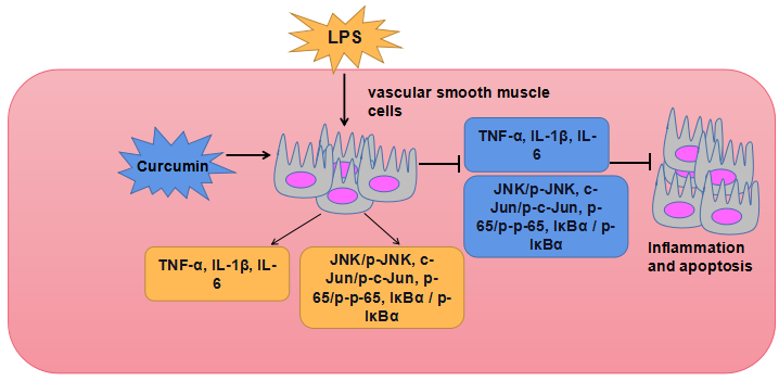
2.1. Cell culture and treatment
Rat cell line VSMCs was cultured in Dulbecco's modified Eagle medium ((DMEM; Gibco BRL,Gaithersburg, MD, USA) containing 10% (v / v) fetal bovine serum (FBS༌Gibco) in 37°C, 5%CO2 atmosphere. Curcumin and LPS are purchased from Sigma-Aldrich (St. Louis, Missouri, USA). Firstly, VSMCs cells were treated with 10 µ M LPS for 12 hours to establish the injury model. Then VSMCs cells were treated with different concentrations of curcumin (0, 50, 100, 150 µ g / mL) for 24 hours.
2.2. Cell viability assay
After digestion, the cells in the logarithmic growth phase were inoculated into 96-well plates at the density of 5 × 10 3/ml for 24 hours, and the old culture was discarded. Curcumin of different concentrations was added to each experimental group for 24 hours. Each experimental group had 4 compound holes, and each well was cultured with MTT solution Sigma-Aldrich (St.Louis, MO) (concentration was 5mg/ml) for 4 hours. The supernatant was carefully discarded, and 200 µ l DMSO, was added to each well to dissolve and dissolve precipitation. The absorbance value was read by the enzyme labeling instrument at the wavelength of 490 nm.
2.3. Apoptosis assay
Cell apoptosis was detected by annexin V-FITC double staining. 24 hours after transfection, the cells were digested by trypsin, collected and inoculated in a 6-well plate, the cell density was adjusted to 2 × 10 6 cells per well, cultured for 24 hours, the supernatant was discarded, pre-cold PBS was washed twice, 1 × Binding buffer was used to suspend cells. The cell suspension was added with 5 µ L annexin V-FITC and 5 µ l PI, After mixing well, the cells were incubated at room temperature for 15 minutes, The apoptotic rate was detected by flow cytometry (Beckman Coulter, Fullerton, CA, USA) within 1 hour, and the data were analyzed by Flow Jo software (tree star, Ashland, or, USA).
2.4. Enzyme-linked immunosorbent assay
VSMCs were seeded in 24 well culture dish and treated with different methods for 24 hours. The culture supernatant was collected. The concentrations of inflammatory cytokines interleukin-6 (IL-6), interleukin-1β (IL-1β) and tumor necrosis factor-α (TNF- α) were measured by ELISA kit (TaKaRa, Dalian).
2.5. Quantitative Real-time PCR
According to the instructions of the manufacturer, total RNA was isolated from VSMCs cells using TRIzol reagent (Invitrogen). Reverse transcription was carried out through multiple transcription kits (Applied Biosystems, Foster, CA, USA). To analyze IL-6, IL-1 β and TNF- α, SYBR green PCR kit (TaKaRa) was used to quantify the messenger RNA (mRNA) levels of IL-6, IL-1 β and TNF- α. β-actin was amplified as a control. The relative expression levels of IL-6, IL-1 β and TNF- α were calculated by the 2−ΔΔCT method.
2.6. Western blot
The total proteins of different groups of cells were extracted by a protein extraction kit. The protein concentration was determined by BCA (Pierce, Appleton, WI, USA). Total proteins were separated by SDS-PAGE electrophoresis with 50 µ g total proteins in each well. After electrophoresis for 2 hours, the membrane was transferred to the PVDF membrane (Millipore, Billerica, Ma, USA) by the wet method. The membrane was sealed with 5% skimmed milk powder for 1 hour, and the primary antibody anti-Bax (ab32503, 1:1000), anti-pro-Caspase3 (ab32150, 1:1000), anti cleaved-Caspase3 (ab32042, 1:1000), anti-IL-6 (ab233706, 1:1000), anti IL-1β (ab216995, 1:1000), anti-TNF-α (ab183218, 1:1000), anti-p65 (ab32536, 1:1000), anti-toll-like receptor 4 (anti-TLR4, ab22048, 1:500), anti-p-p65 (ab31624, 1:1000), anti-IκBα (ab109300, 1:1000), anti-JNK (ab176645, 1:1000), anti-p-JNK (ab176645, 1:1000), anti-c-Jun (ab40766, 1:1000) and anti-p-c-Jun (ab32385, 1:1000), anti-β-actin(ab8226, 1:2000), all purchased from Abcam (Cambridge, UK) was incubated at 4 ℃. Overnight, In the next morning, TBST was used to rinse the membrane, horseradish peroxidase-labeled second antibody was added and incubated at 37 ℃ for 1 h. ECL was used to develop and save the image. The optical density value was measured by quantity one.
2.7. Statistical analysis
The data of this study were expressed by mean ±standard deviation (SD), and the statistical software SPSS 19.0 was used (IBM Analytics, New York, USA), P < 0.05was considered to be a statistically significant result. All experiments are repeated at least three times.