3.1 Working principle of the developed DLS immunosensor for NT-proBNP
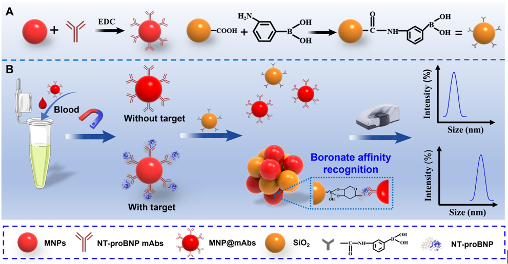
Scheme 1 describes the working principle of the developed DLS immunosensor for the quantitative detection of NT-proBNP, wherein MNP@mAb was employed for magnetic enrichment of target analytes and DLS signal transduction, and SiO2@PBA was designed as crosslinkers to amplify the crosslinking aggregation of MNPs. When NT-proBNP was present in the sample solution, target glycoprotein was selectively captured by the MNP@mAb to form the immunocomplex of MNP@mAb-NT-proBNP. After washing to remove the supernatant, the immunocomplex was re-suspended in PB solution containing boronic acid crosslinkers, thereby inducing the MNP aggregation by the selective boronic acid ligand–cis-diol recognition between the SiO2@PBA and the glycoprotein. With the crosslinking aggregation of MNPs, the DH of the solution will remarkably increase, which can be readily measured by DLS. Specifically, when the content of NT-proBNP is more, the MNP aggregate is larger, and the DH of the solution is greater. By contrast, no obvious aggregation of MNPs was observed when the target NT-proBNP was absent, thus resulting in negligible changes in the DH. Therefore, the quantitative detection of glycoproteins in unknown samples can be achieved by recording the variation in the DH of MNPs.
3.2 Synthesis and characterization of MNP@mAb and SiO2@PBA
The MNP@mAb conjugates were prepared through the formation of peptide linkage between the carboxyl group of MNPs and the amino group of anti-NT-proBNP mAb in the presence of EDC. The successful construction of the MNP@mAb was confirmed by TEM and DLS. As shown in Figure 1A, the MNPs show uniform morphology and good monodispersity before and after conjugated with mAb. DLS measurement indicates an obvious increase in the DH of MNPs from 139 nm to 149.7 nm after the conjugation of mAbs (Figure 1B). Figure 1C exhibits the MNP@mAb has higher zeta potential of −36.7 mV than that of unmodified MNPs (−56.1 mV). These results suggest the successful conjugation of MNPs with mAbs. As shown in Scheme 1A, the SiO2@PBA conjugates were synthesized by a similar EDC-assisted covalent coupling method. The successful modification of PBA molecules on the surface of SiO2 was verified using TEM, DLS and FTIR. Figure 1D and Figure S1 reveals that no obvious changes in the morphology and monodispersity of SiO2 were observed after modified with the PBA. Figure 1E shows that the size of SiO2 increased from 114.4 nm to 124 nm with the zeta potential reduced from −15.0 mV to −31.0 mV when PBA molecules were modified onto the surface of SiO2. Further FTIR analysis of SiO2@PBA was compared with SiO2. As shown in Figure 1F, FTIR spectra of SiO2 and SiO2@PBA present two characteristic peaks at 1057 cm−1 and 3273 cm−1, which correspond to the Si–O band and −OH, respectively [27]. In addition, the FTIR peaks of SiO2@PBA at 1633 cm−1, 1541 cm−1 and 1421 cm−1 correspond to the stretching vibration of a benzene ring skeleton, the peptide bond and −B(OH)2, respectively, which were not observed in SiO2 alone [28]. These findings prove that PBA molecules were successfully modified onto the surface of SiO2.
3.3 Confirmation of feasibility of the developed DLS immunosensor for NT-proBNP
The feasibility of the developed DLS immunosensor for the quantitative analysis of NT-proBNP was verified by conducting a series of control experiments, including (1) MNP@mAb, (2) MNP@mAb + NT-proBNP (20 pg mL−1), (3) MNP@mAb + SiO2@PBA, and (4) MNP@mAb + NT-proBNP (20 pg mL−1) + SiO2@PBA. The formation of MNP aggreates caused by target NT-proBNP was monitored by DLS and TEM. Results in Figure 2A and Figure S1 present that only the co-occurrences of target NT-proBNP and SiO2@PBA (denoted as MNP@mAb + NT-proBNP (20 pg mL−1) + SiO2@PBA) can result in a significant increase in the DH of MNP@mAb from 147 nm to 351 nm. By contrast, two other groups, including MNP@mAb + NT-proBNP and MNP@mAb + SiO2@PBA, show negligible changes in the DH compared with MNP@mAb alone. The reason for this phenomenon is that the presence of target NT-proBNP can specifically induce the MNP aggregation caused by SiO2@PBA, which are well demonstrated by SEM and TEM imaging (Figure 2B-C). The formation of MNP aggreates caused by the SiO2@PBA was further confirmed by energy dispersive spectroscopy (EDS) mapping analysis (Figure 2D) with the coexistence of Si and Fe in the aggregates. These observations verify the feasibility of the proposed boronate affinity amplified DLS immunosensor for ultrasensitively and specifically targeting NT-proBNP.
3.4 Optimization of experimental conditions
To achieve the best analytical performance of the developed DLS immunosensor, several key parameters, such as the pH value, EDC amount, and labelled amount of mAbs for the preparation of MNP@mAb; the used amount of MNP@mAb for each test; the immunoreaction time of MNP@mAb and target NT-proBNP; the used amount of SiO2@PBA, pH value and incubation time for boronate affinity reaction between NT-proBNP and SiO2@PBA, were systematically studied. The optimized experimental conditions were evaluated by detecting the largest DH change using DLS. The results in Figure 3A-C indicate the optimal combinations of pH value, EDC amount, and labelled amount of mAbs for the preparation of MNP@mAb were 7.5, 25 µg mL−1, and 100 µg mg−1, respectively, wherein the MNP@mAb maintains the best bioactivities for the recognition and capture of target NT-proBNP, thus giving the maximal DH values. Figure 3D show that the DH gradually increased with increasing the used amount of MNP@mAb from 1.2 µg to 2.4 µg per test, and then obviously decreased with the further increase of MNP@mAb, which may be due to the presence of excess MNP@mAb to cause a reduction in the aggregate formation and size. The immunoreaction time between the MNP@mAb and the target NT-proBNP was further investigated to ensure the high sensitivity and reproducibility. Figure 3E shows that 5 min of immunoreaction time was necessary to result in the maximum DH with high reproducibility. In addition, to further maximize the variation in the DH, the boronate affinity recognition between the target glycoprotein and the boracic acid crosslinker is critical to control the crosslinking aggregation of MNPs for amplifying the DLS signal and improving the sensitivity. Figure 3F displays the effect of pH ranged from 7 to 9 on the MNP aggregation, and the results show the greatest DH value at pH 7.5. With increasing the amount of SiO2@PBA from 0.032 µg mL−1 to 4 µg mL−1, the DH value increased gradually from 224.7 nm to 259.6 nm (Figure 3G). However, the DH value decreased obviously as the concentration of crosslinking agents continued to increase to 20 µg mL−1. The possible reason is that the excess crosslinker could block the remaining cis-diol sites on the glycoprotein molecules and in turn inhibit the MNP aggregation, thus giving rise to a decreased DH. Figure 3H shows that at all NT-proBNP concentrations, the DH value reached a constant after 5 min reaction of the glycoprotein and the SiO2@PBA, suggesting 5 min was enough to allow sensitive and reproducible DLS signal transduction for reliable quantitative analysis.
3.5 Performance evaluation of the amplified DLS immunosensor
The signal response of this DLS immunosensor against different concentrations of NT-proBNP was examined under the developed conditions. As shown in Figure 4A, the DH value gradually increased with the NT-proBNP concentration ranging from 0.012 pg mL−1 to 1100 pg mL−1. Figure 4B shows an excellent linearity between the DH value and the logarithm of the NT-proBNP concentration (0.012-100 pg mL−1). The linear regression equation is described as: y = 21.885lnx + 255.26, with the correlation coefficient of 0.9858. The limit of detection was 0.0074 pg mL−1 (S/N=3), which is much lower than other previously reported immunoassays for NT-proBNP detection (Table 1), demonstrating that our immunosensor has better sensitivity. Notably, the total time to complete a test is less than 20 min, which is comparable to that of the most widely used POC device of lateral flow assay and obviously shorter than other reported immunoassays. The ultrahigh sensitivity and fast response time are ascribed to the boronate affinity-based multivalent recognition between crosslinkers and target glycoproteins and the magnetic enrichment of targets from the complex sample. Additionally, an obvious hook effect was observed at the NT-proBNP concentration of over 200 pg mL−1, indicating that additional sample dilution was needed to ensure accurate quantification of NT-probNP and avoid false results when the concentration of NT-probNP exceeds the hook point.
The specificity of this developed DLS immunosensor was characterized by using a series of common interfering glycoproteins, including carcinoembryonic antigen (CEA), alpha fetoprotein (AFP), human chorionic gonadotropin (HCG), and hepatitis B virus antigen (HBsAg), as well as monosaccharides such as glucose (Glc), galactose (Gal), fucose (Fuc), and nacetylneuraminic acid (Neu5Ac). Figure 4C indicated that only NT-proBNP can provide an apparent signal value, while other interferents show negligible changes compared with the negative control. These results indicate that the designed DLS immunosensor can specifically distinguish target glycoprotein from other interfering substances because of the specific immunorecognition between NT-proBNP and its captured antibody.
The accuracy and precision of the proposed DLS immunosensor for the quantitative detection of NT-proBNP were further investigated by measuring the intra- and inter-assay recoveries. As indicated in Table S1, the intra-assay recoveries ranged from 89.8–101.13% with the coefficient of variation (CV) of 0.32–10.77%, while the inter-assay recoveries varied from 80.81–104.11% with the CV of 3.91–7.87%. These results indicate an acceptable accuracy and precision for quantitative determination of NT-proBNP in complex samples.
The reliability and practicability of this DLS immunosensor in actual samples were demonstrated by the detection of NT-proBNP in human serum. Forty NT-proBNP-positive serum samples collected from the First Affiliated Hospital of Nanchang University were simultaneously analyzed by the developed immunosensor and the clinical routine timed-resolved fluoroimmunoassay (TRFIA). For our method, 1 µL of serum samples were diluted with 199 µL of 0.01 M PB solution (pH 7.4) and then quantified, while the analysis of NT-proBNP using the TRFIA was performed according to the manufacturer’s instructions. The test results obtained by these two approaches were then compared. As indicated in Table S2, among these 36 samples, 34 samples were tested with NT-proBNP as detected by our proposed DLS immunosensor method. By contrast, 30 samples were tested NT-proBNP positive by the TRFIA method. These results showed that compared with the routinely used TRFIA, the newly developed DLS immunosensor can detect lower concentrations NT-proBNP, which is attributed to its higher sensitivity (LOD, 1.48 pg mL−1) than that of the TRFIA method (70, pg mL−1). In addition, 2 samples were simultaneously detected NT-proBNP negative by these two methods. Figure 4D and Figure S3 exhibit that the detection results of the proposed method are broadly in line with those of TRFIA with a good linear correlation of 0.9745, proving the feasibility of the amplified DLS immunosensor for real-world applications in complex sample matrix.