2.1. Subjects
This is retrospective case study was exempted from institutional review board of Nanoori Hospital, Seoul, Republic of Korea. The informed consent was obtained from all patients participated in study. We studied 18 consecutive patients with 20 levels of lumbar degenerative disease; (6 males, 12 females; Mean age 63.71 years) who underwent Endoscopic Transforaminal Lumbar Interbody Fusion(TLIF) through the Posterior Paraspinal Approach at a spine specialty hospital between august 2018 and January 2019. Inclusion criteria was degenerative grade 1 spondylolisthesis, lumbar central canal stenosis with foraminal stenosis and segmental instability confirmed on dynamic radiograph. Spondylolisthesis grade 2 and more and lumbar canal stenosis with more than 50% loss of disc height are excluded from study due to difficulty to be managed with endoscopic fusion.
2.2. Surgical procedure
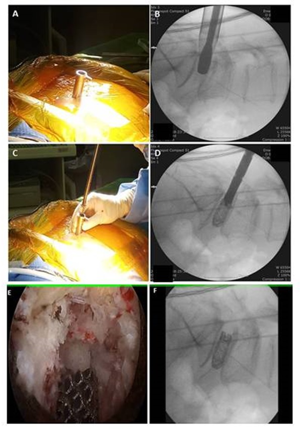
Procedure done under general anaesthesia. Patient placed in prone position on radiolucent table over Wilson's frame. Entry point is located under fluoroscopy guidance. Target point is ipsilateral facet at the level and it is approached through conventional Wiltse's approach between multifidus and longissimus muscle. Usually interval located about 3cm lateral to the midline. A longitudinal skin incision of approximately 1cm in length was made 4cm lateral to the midline on affected side. It facilitates the medial trajectory of working cannula and cage into the disc space. Serial dilators introduced through the interval and finally working channel introduced along with endoscope. For initial dissection we use working cannula with outer diameter of 13.7 mm and bevelled tip, the endoscope has 15 degree viewing angle, outer diameter of 10 mm, working channel diameter of 6 mm and working length 125 mm. ILESSYS-DeltaR (Joimax Gmbh, Germany).
The soft tissue over facet joint is dissected using radiofrequency ablator to expose inferior articular process(IAP) of superior vertebra. Endoscopic drill is used to hollow out the groove at IAP in inverted "L" fashion and its resected with bone cutter and forceps. IAP harvested in total to use as a autologus graft later. Underlying superior articular process(SAP) burred with drill to expose foraminal part of deep layer of ligamentum flavum. Central and contra lateral decompression can conveniently achieved though the same interval by simply tilting the endoscope. [figure 1] Haemostasis and annulotomy done with radiofrequency probe (Elliquence, New York, USA). Disc material removed with forceps. End plate preparation is most important step of the procedure and advantage of endoscopic spine surgery is meticulous end plate preparation can be done under direct vision without damaging it. End plate cartilage is removed with dissector and curettes to prepare fusion bed in order to protect traversing root bevel tip of cannula is rotated from superior to medial direction and inserted into the disc space through the annulotomy site. Autologus graft mixed with allograft is tamped into the disc space under image fluoroscopy guidance. Single large 3D printed cage (GENQSS, GS Medical, Republic of korea) with demineralised bone matrix(DBM) is inserted as oblique as possible and final position of cage is checked under both fluoroscope as well as endoscope. Working cannula is withdrawn keeping suction drain in situ. [picture 1]
After completion of interbody fusion procedure construct is stabilized with percutaneous pedicle screw fixation through same incision on ipsilateral side. Same procedure is repeated on contra lateral side through 2 separate incisions and rods are inserted with compression in routine fashion. If required pedicle screw fixation is augmented with bone cement. [video]
2.3 Outcome evaluation
a) Clinical evaluation: Demographic data collected and all 18 patients were clinically evaluated on the basis of the Visual Analogue Scale(VAS) score for the back, Oswestry Disability Index(ODI) and MacNab's criteria preoperatively, postoperatively and final follow up (minimum 6 months). Completeness of decompression was documented with postoperative magnetic resonance imaging(MRI). Patients are also assessed for any intra or post operative complications and recurrence of symptoms.
b) Radiological evaluation:
The radiological assessment was performed by independent observer who was blinded to the patient's clinical information. Plain radiograph used to evaluate disc height(DH), lumbar lordosis angle(LLA) and segmental lordosis angle(SLA) pre and post operatively.
The fusion status was assessed using computed tomography(CT) at 6 months and 1 year as recommended by CT protocol. [5] Bony fusion was defined when there is continuous contact of trabecular bone between upper and lower endplates of fusion segments. Osteolysis at the margins of fusion devices (pedicle screw), cystic changes within endplates adjacent to the cage, linear defect in trabecular bone bridge of fusion were considered features of delayed or failed fusion.
to evaluate fusion mass, CT scan done with axial slices parallel to the end plates of fused segment. Percentage of fusion mass surface area is calculated on CT images in axial cut using software (Infinitt, inc, Seoul, Korea) in following steps -
- The surface area of fusion mass was measured at mid disc level from the region of interest (ROI)(surface area of cage is also included as we have used 3D-printed cage with osteo-integration property; surface area of osteophytes at end plate excluded)
- the surface area of upper and lower end plates measured from the slices going through or close to the end plates
- average of the upper and lower end plates surface area calculated
percentage of fusion mass surface area calculated with the formula:
percentage = surface area of fusion mass/average end plate surface area X 100 [figure 2]
2.4 Statistical analysis
Clinical data was analyzed with SPSS version 18 statistical analysis software (IBM corporation, New York). The continuous variables were expressed as mean and standard deviation(SD). The paired t test is used for comparison of preoperative and postoperative VAS, ODI scores; LLA, SLA and DH values. A value of (p < 0.01) considered significant.